
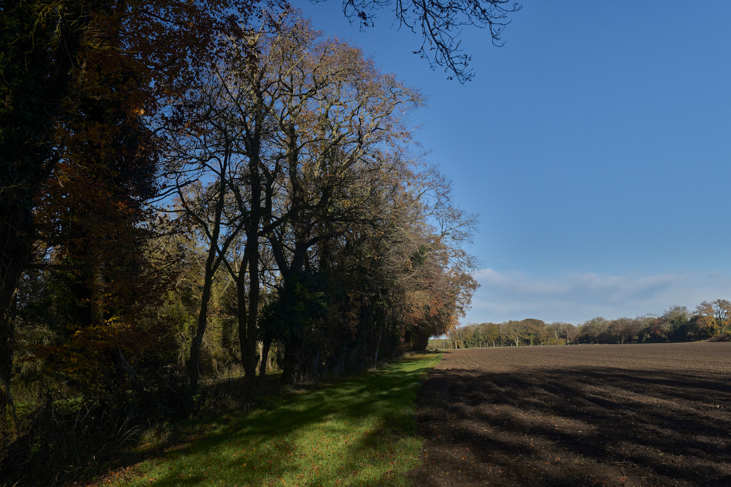
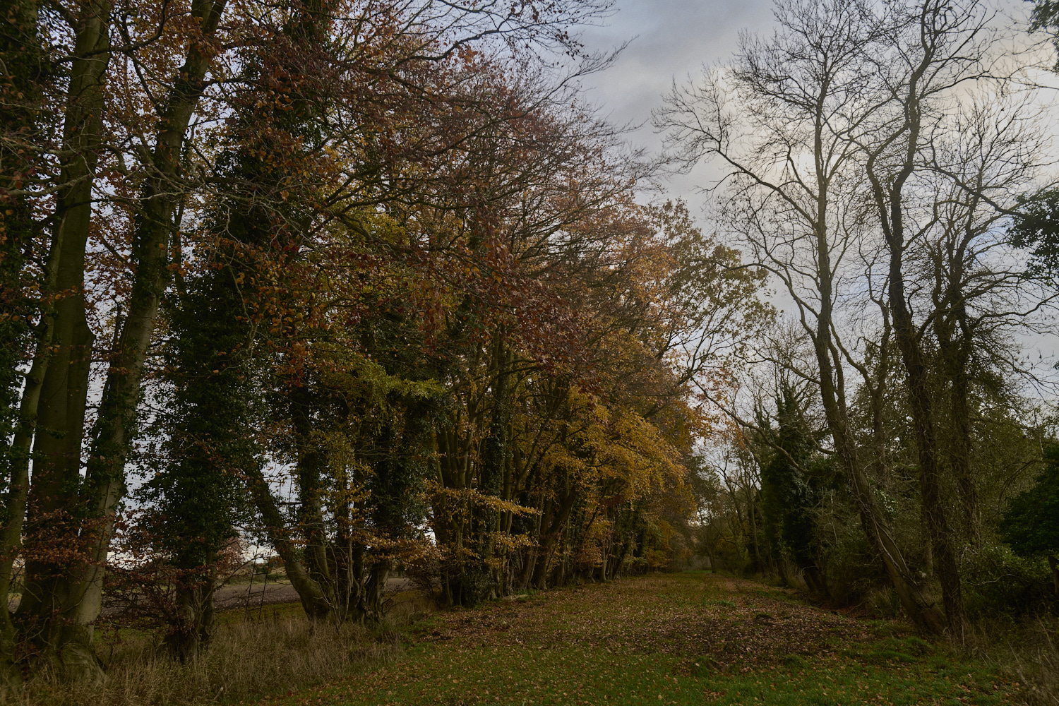
East Harling
Something Natural Surroundings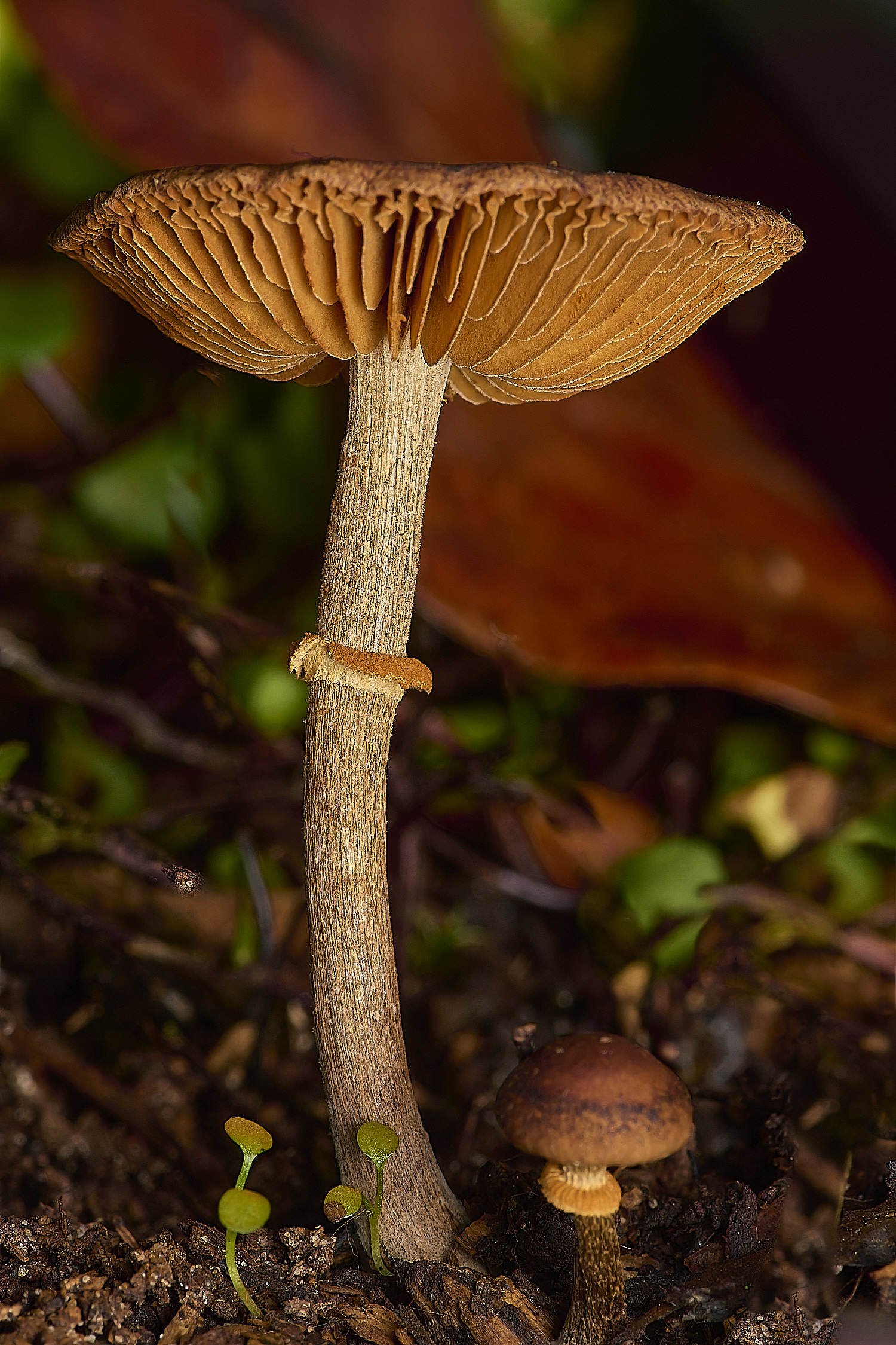
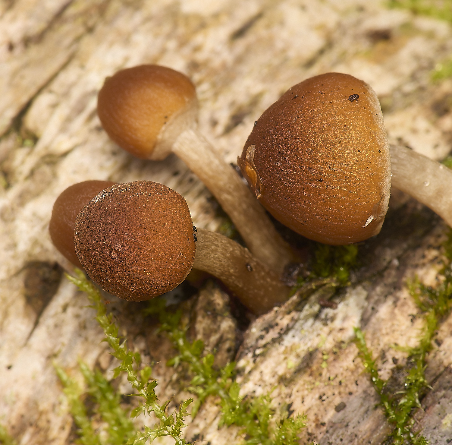
Common Conecap (Pholiotina rugosa)
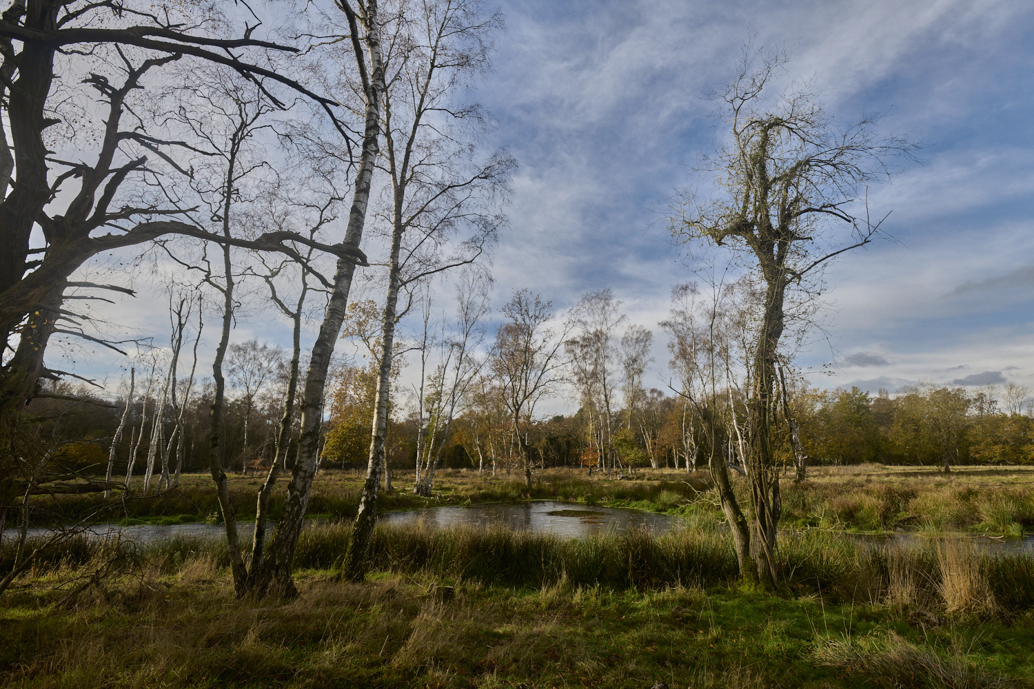
East Harling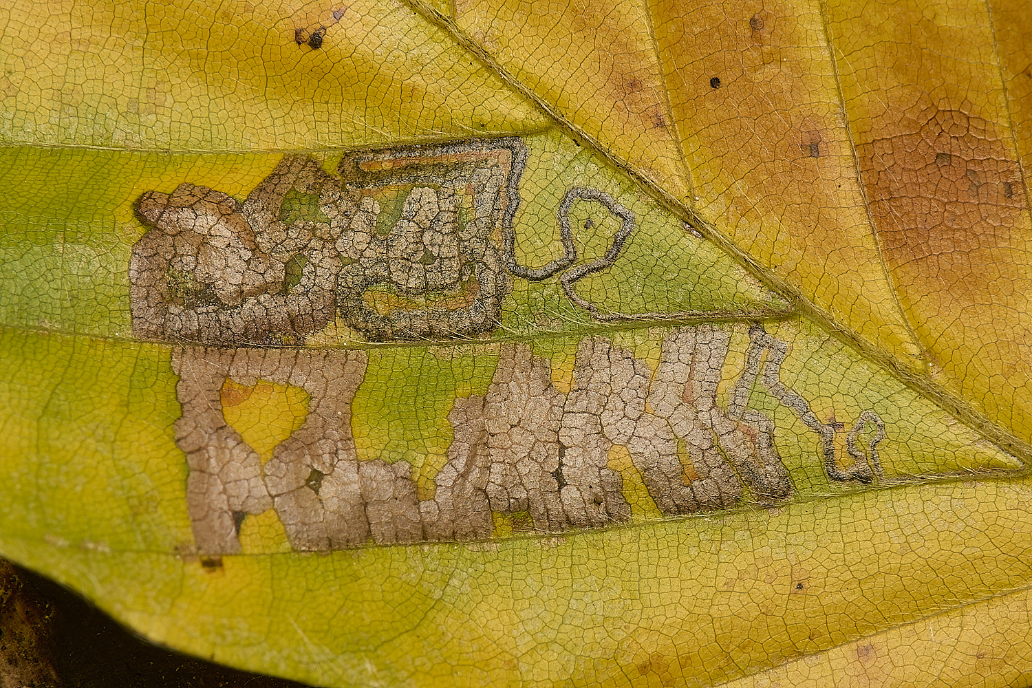
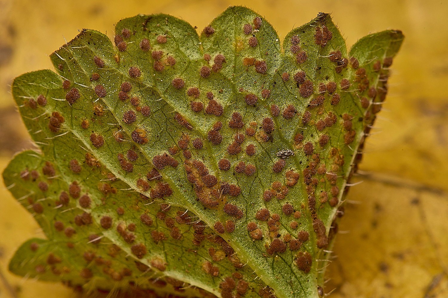
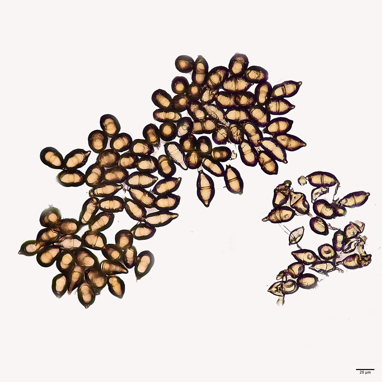
Small Beech Dot (Stigmella tityrella) on a Beech (Fagus sylvatica) leaf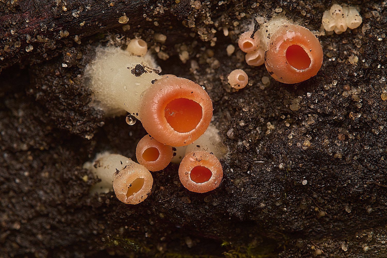
Young Scarlet Elf Cups (Sarcoscypha austriaca)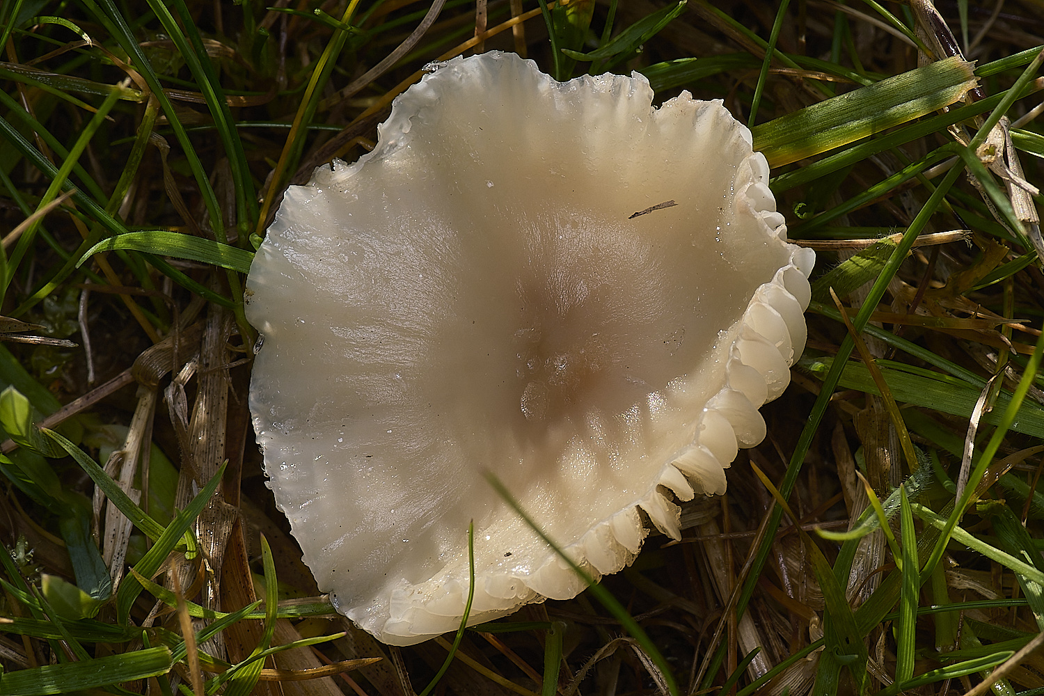
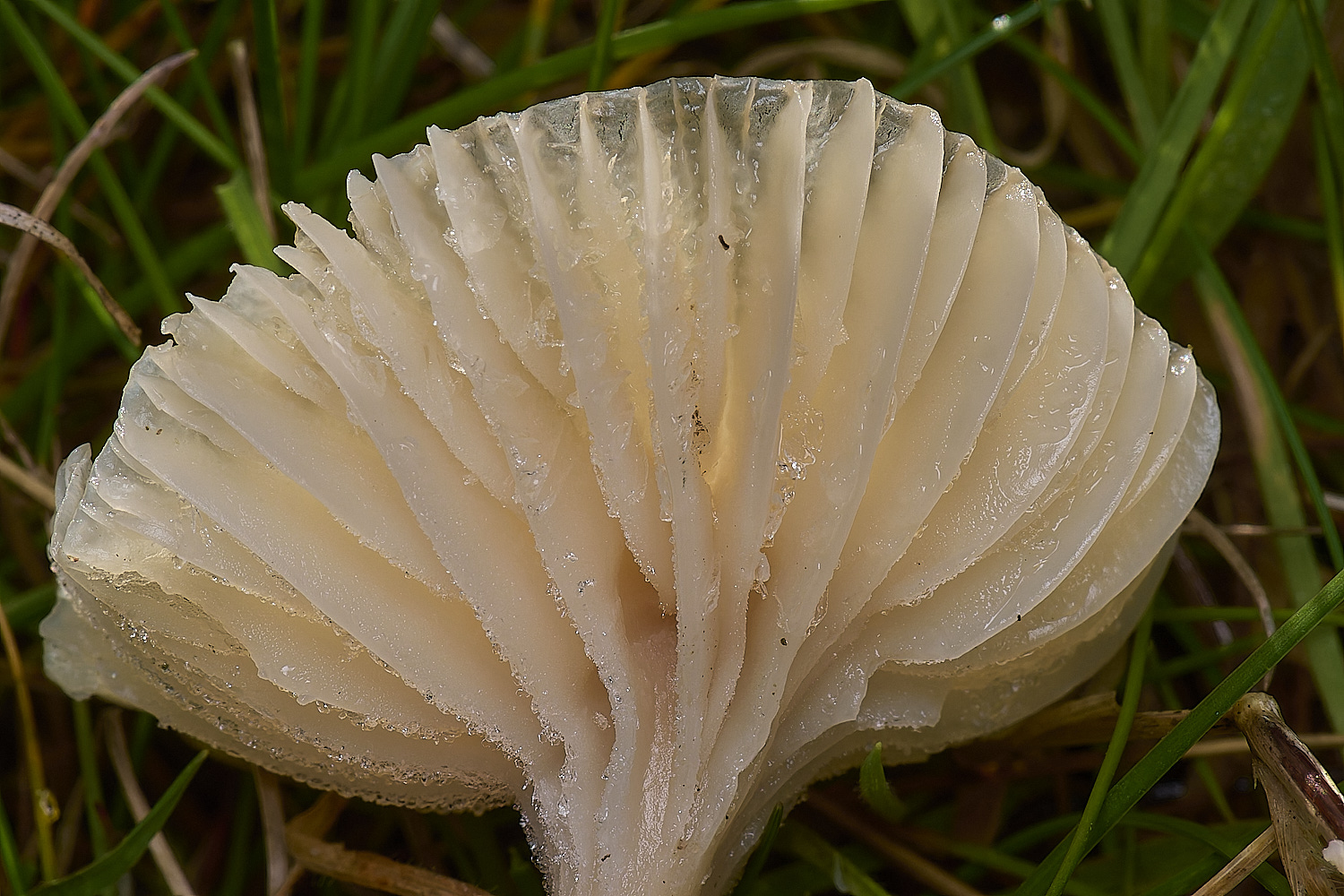
Snowy Waxcap (Cuphophyllus virgineus)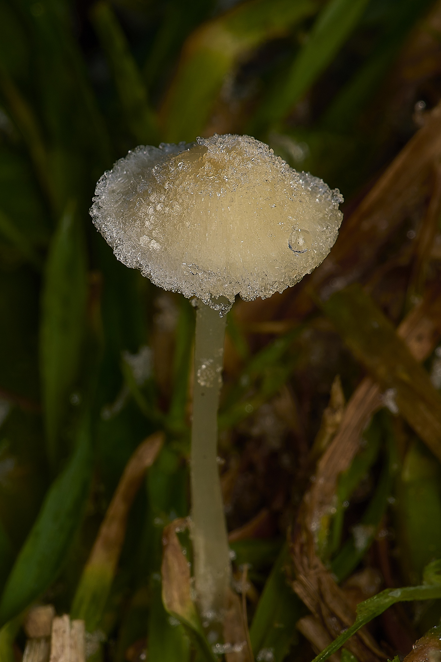
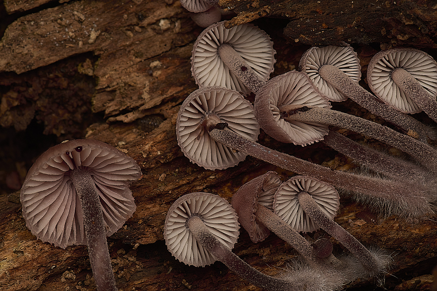
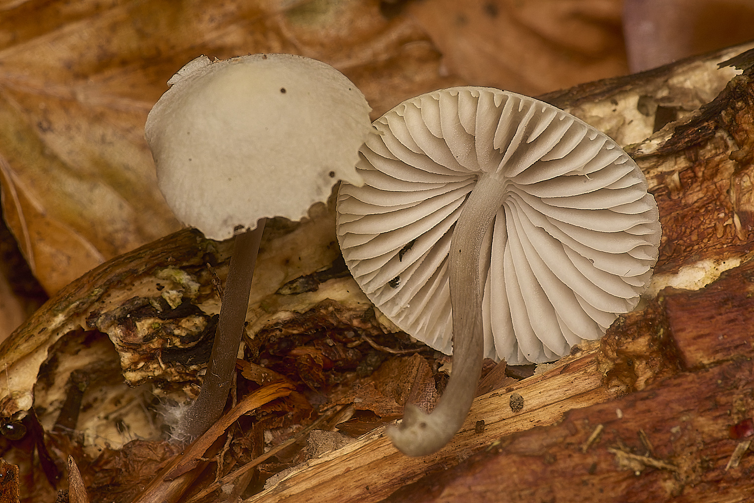
Ivory Bonnet (Mycena flavoalba))
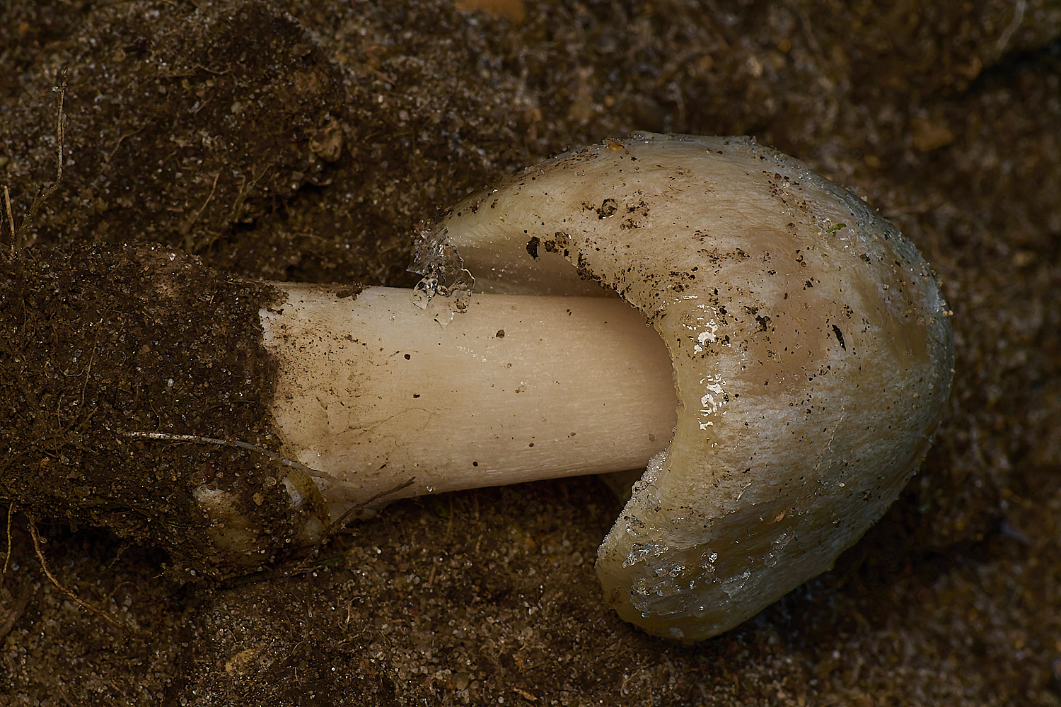
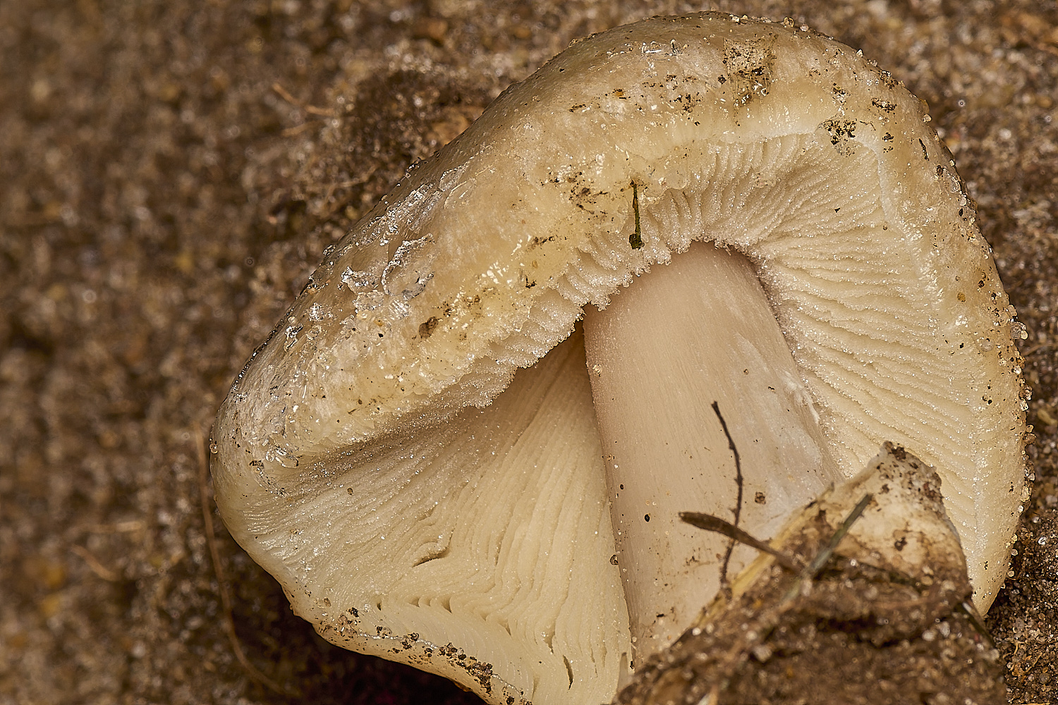
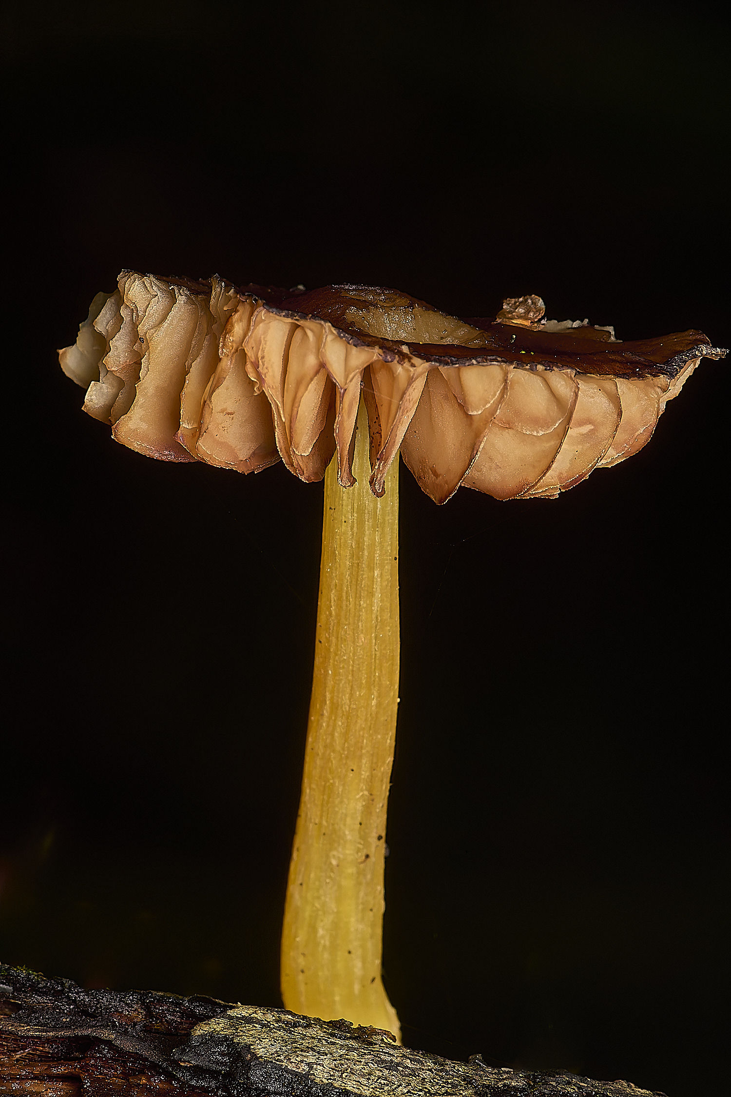
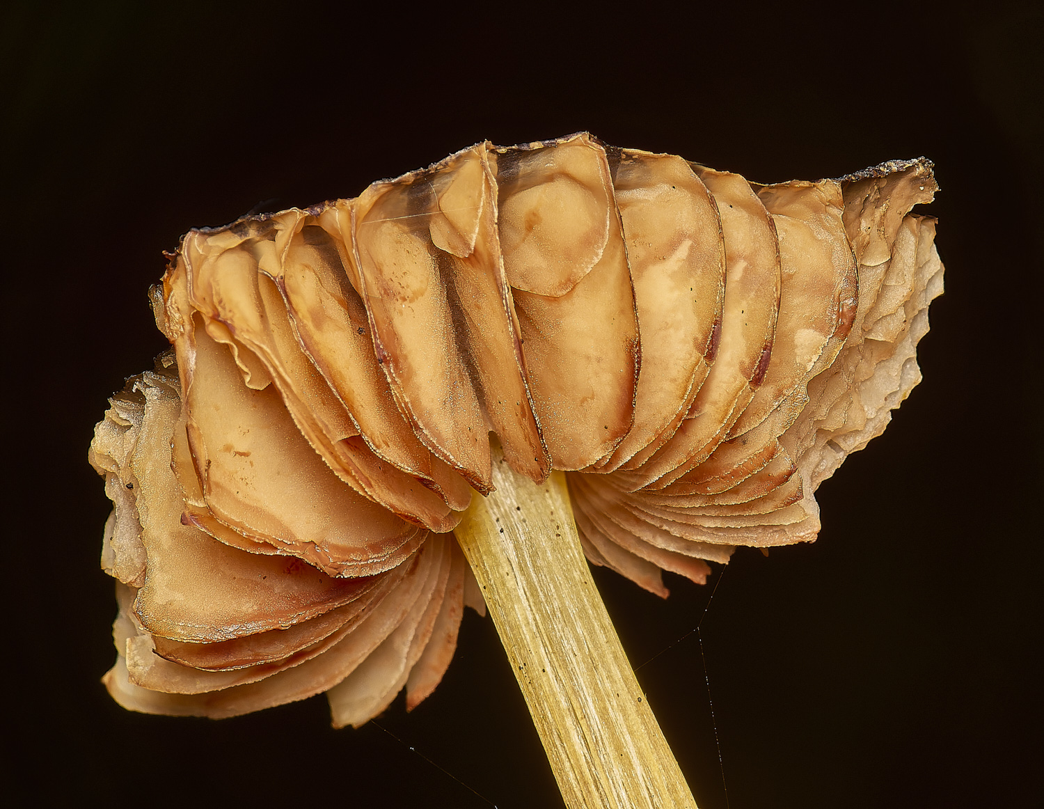
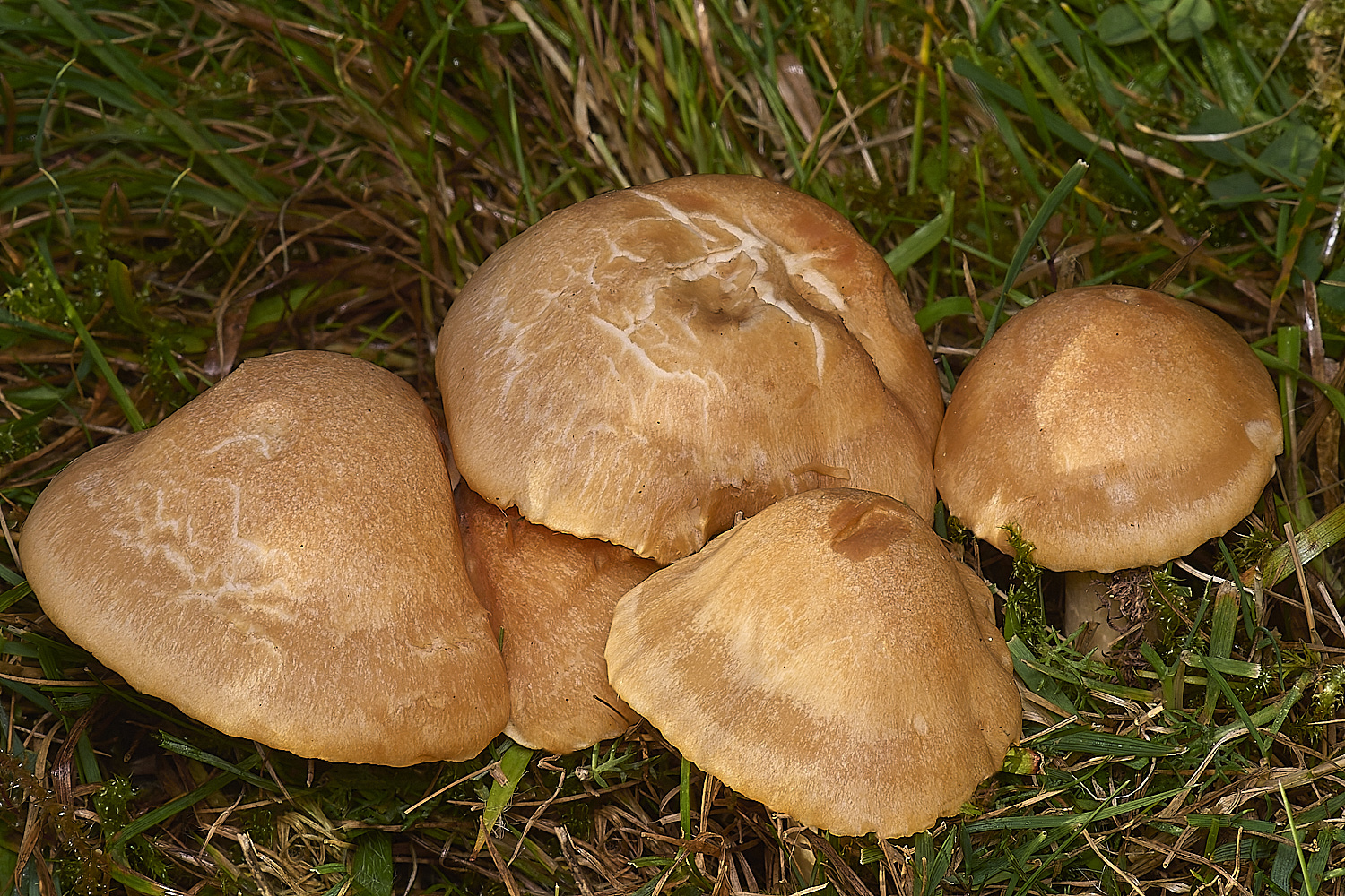
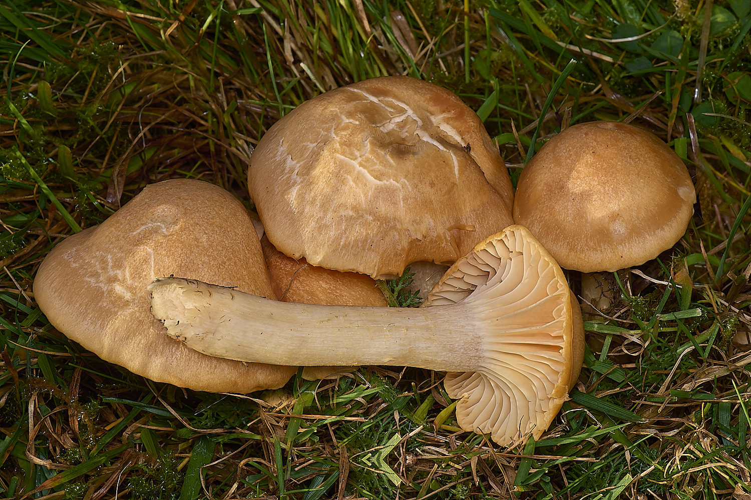
Stubble Rosegill (Volvopluteus gloicephalus)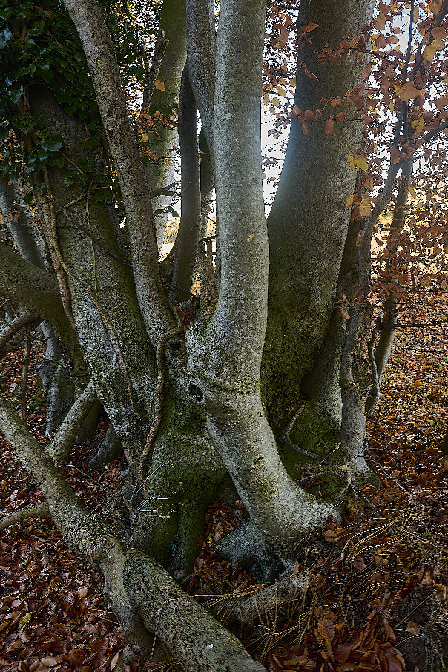
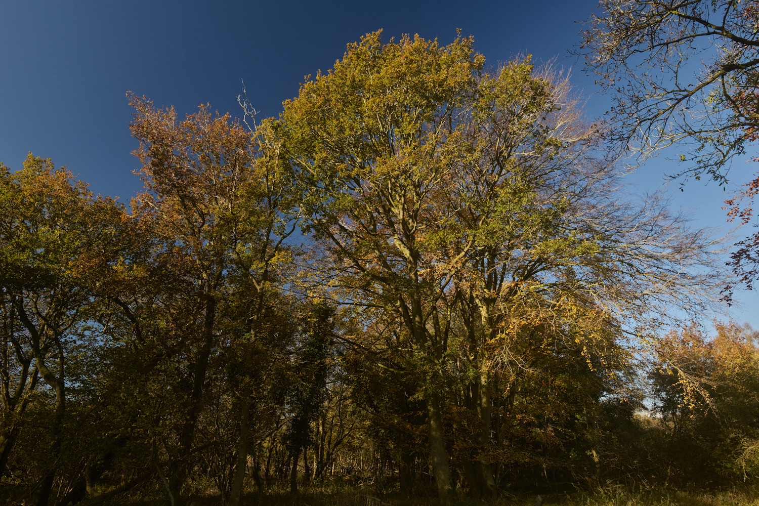
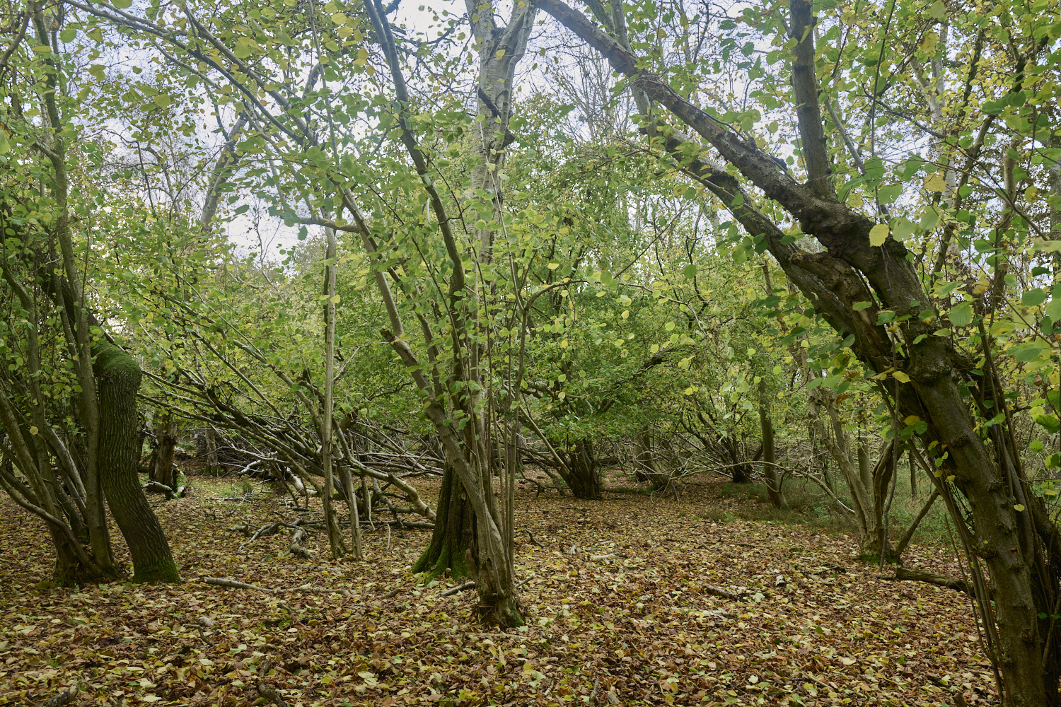
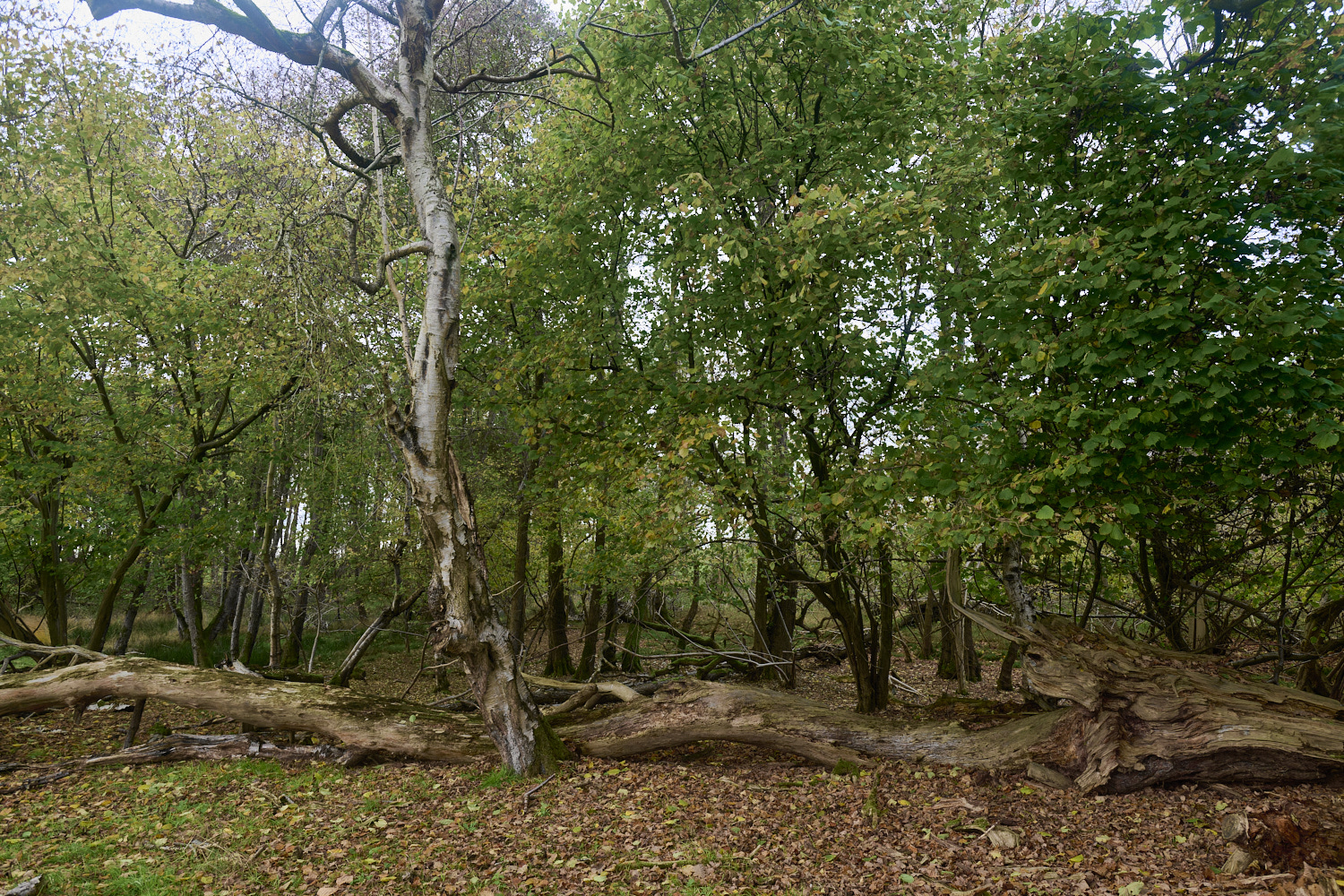
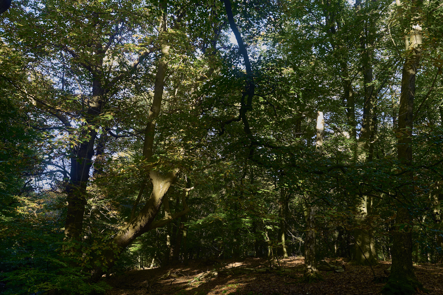
Strong grey Beech (Fagus sylvatica) trunks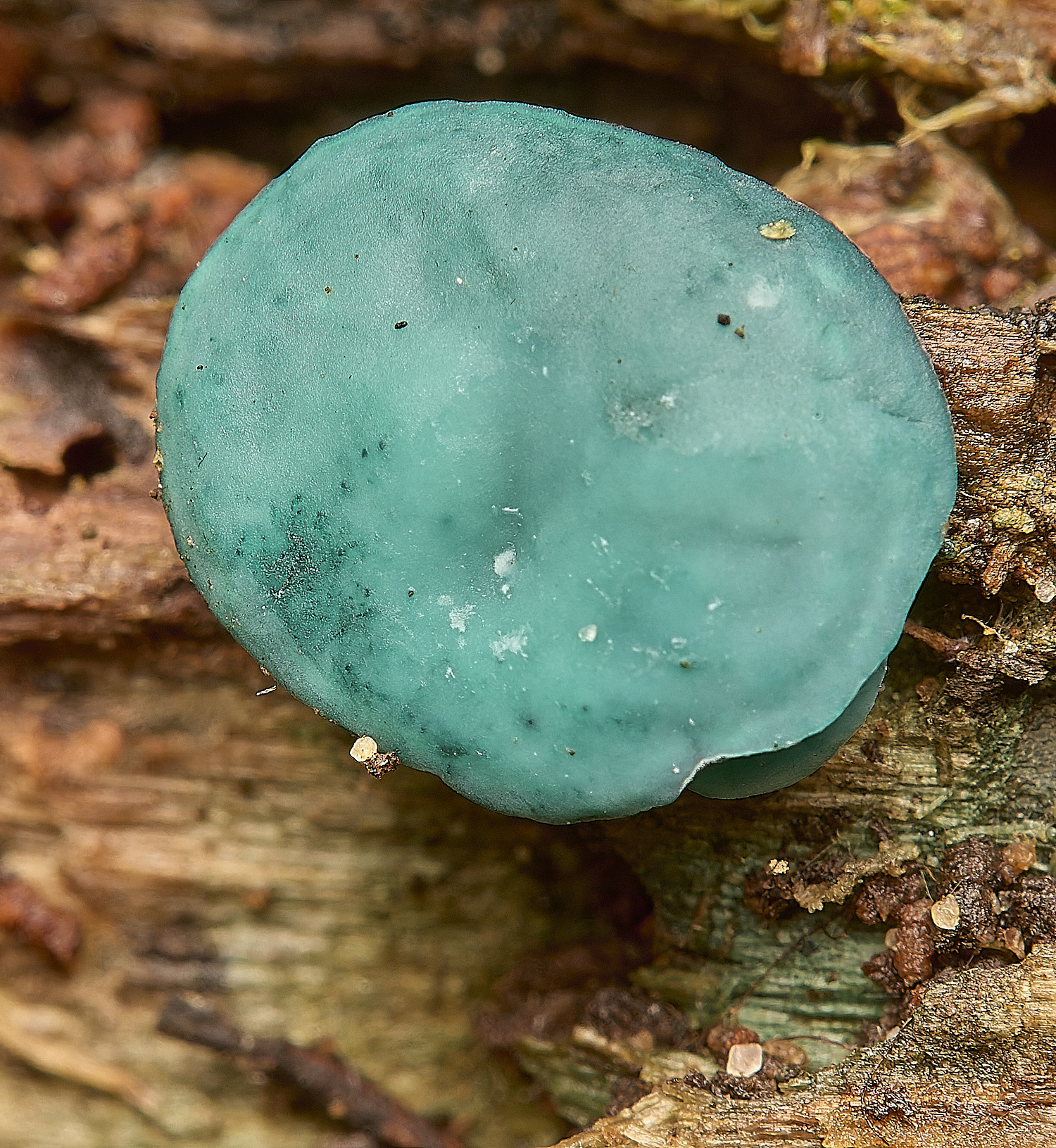
Green Elf Cup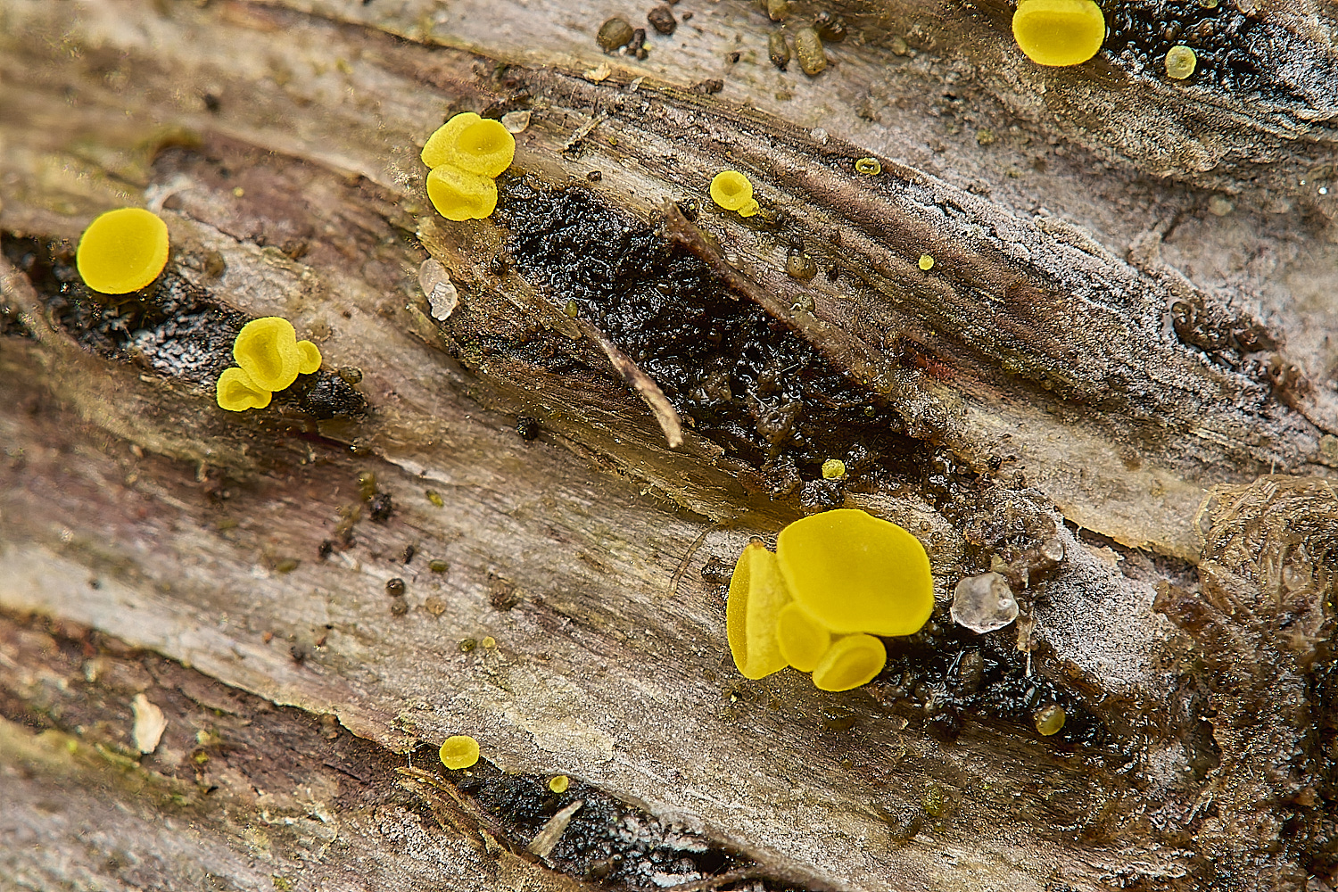
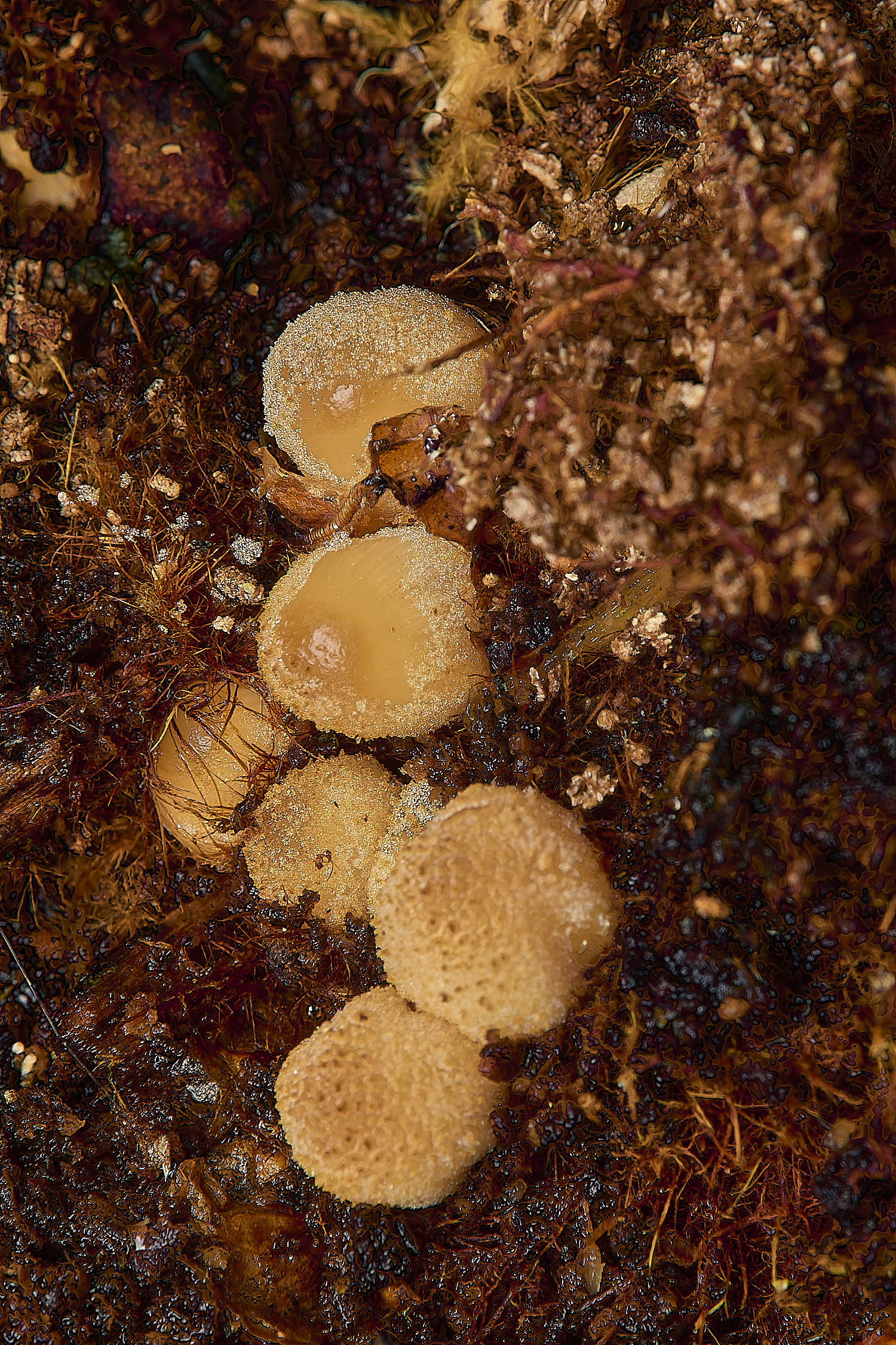
Sulphur Disco (Bisporella sulfurina)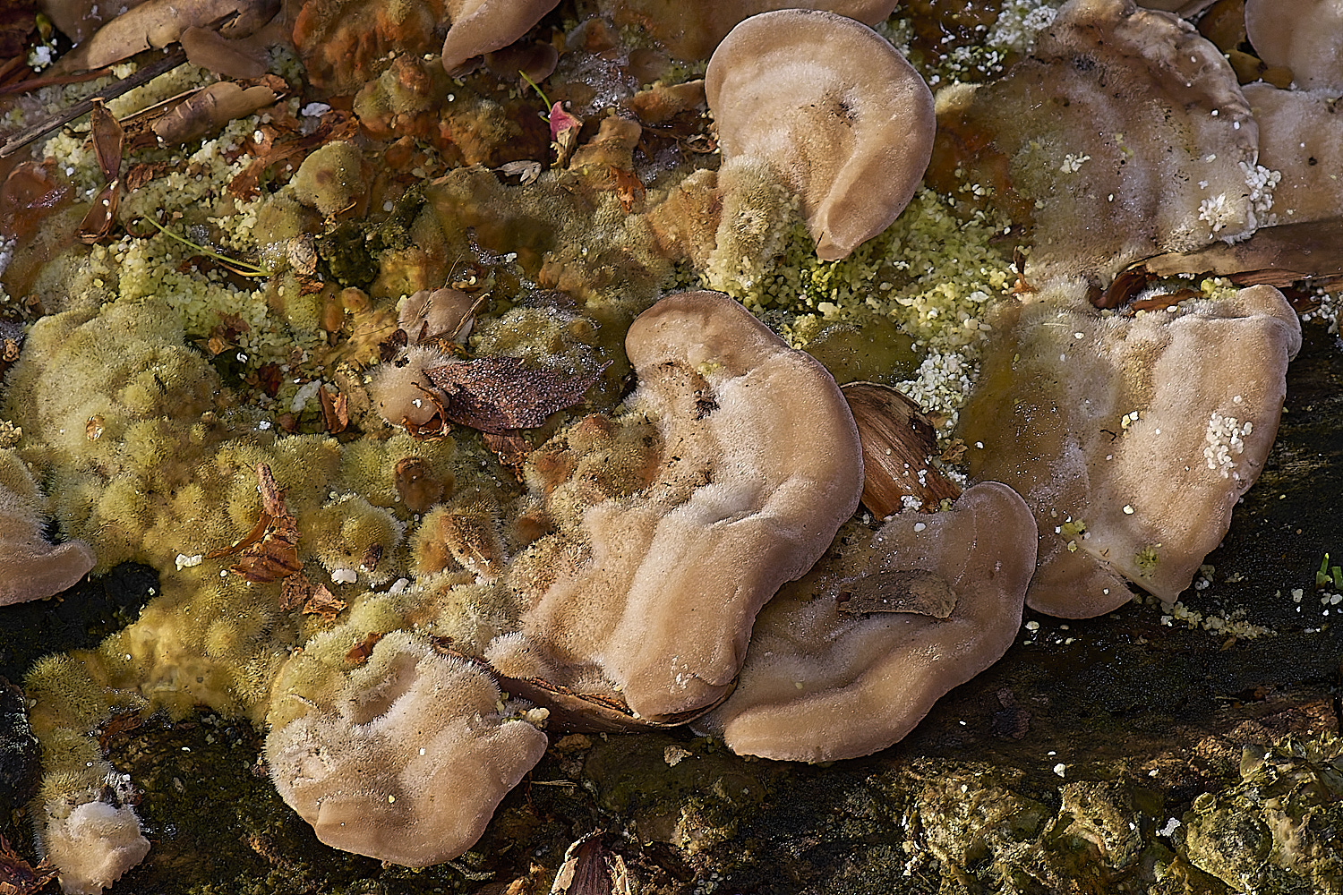
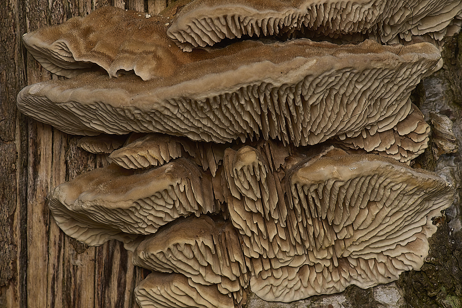
Birch Mazegill (Lenzites betulinus)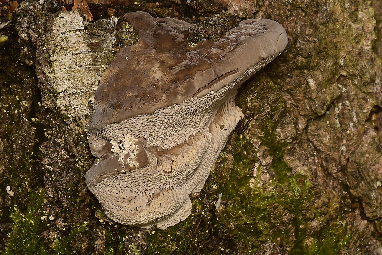
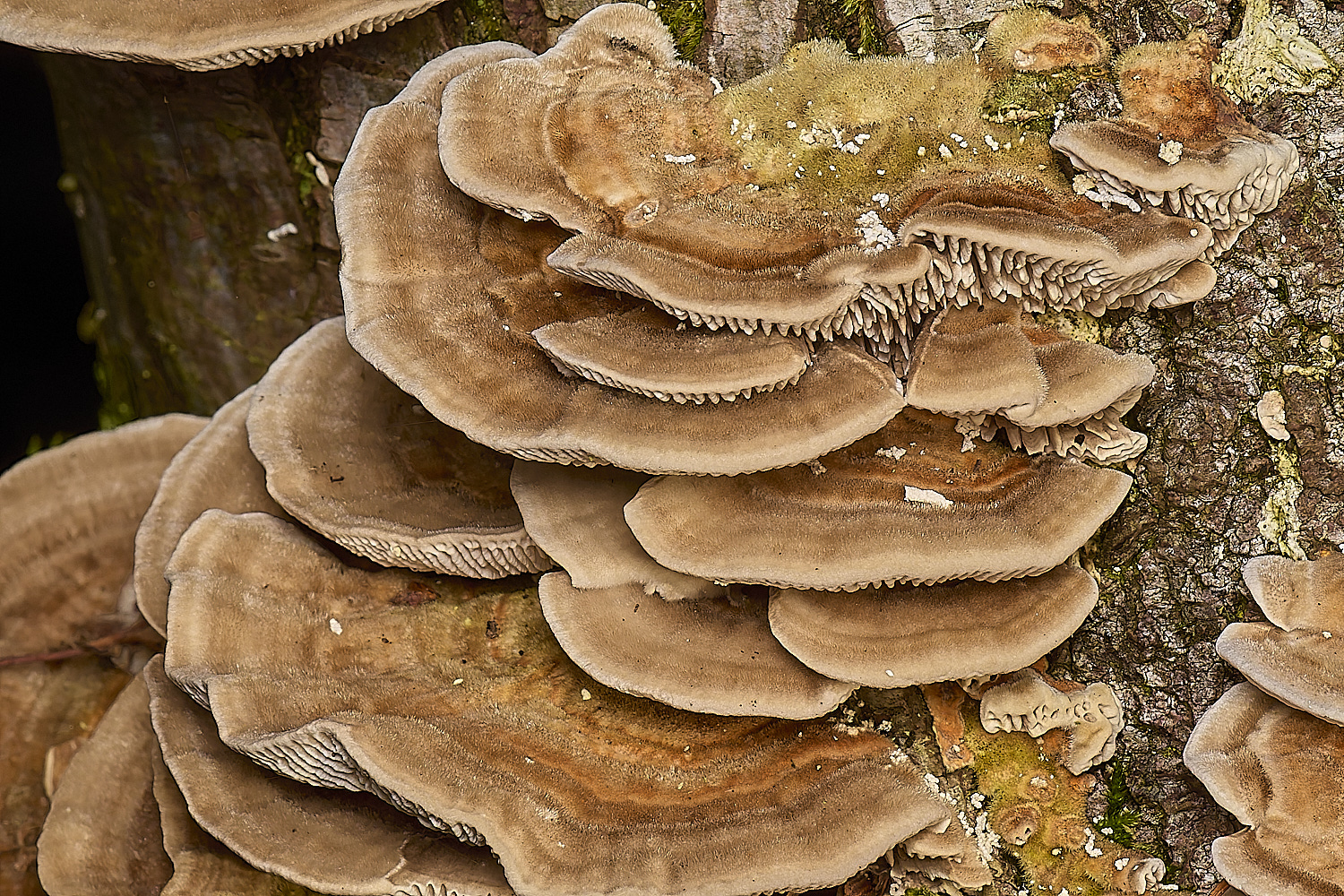
Lumpy Bracket (Trametes gibbosa)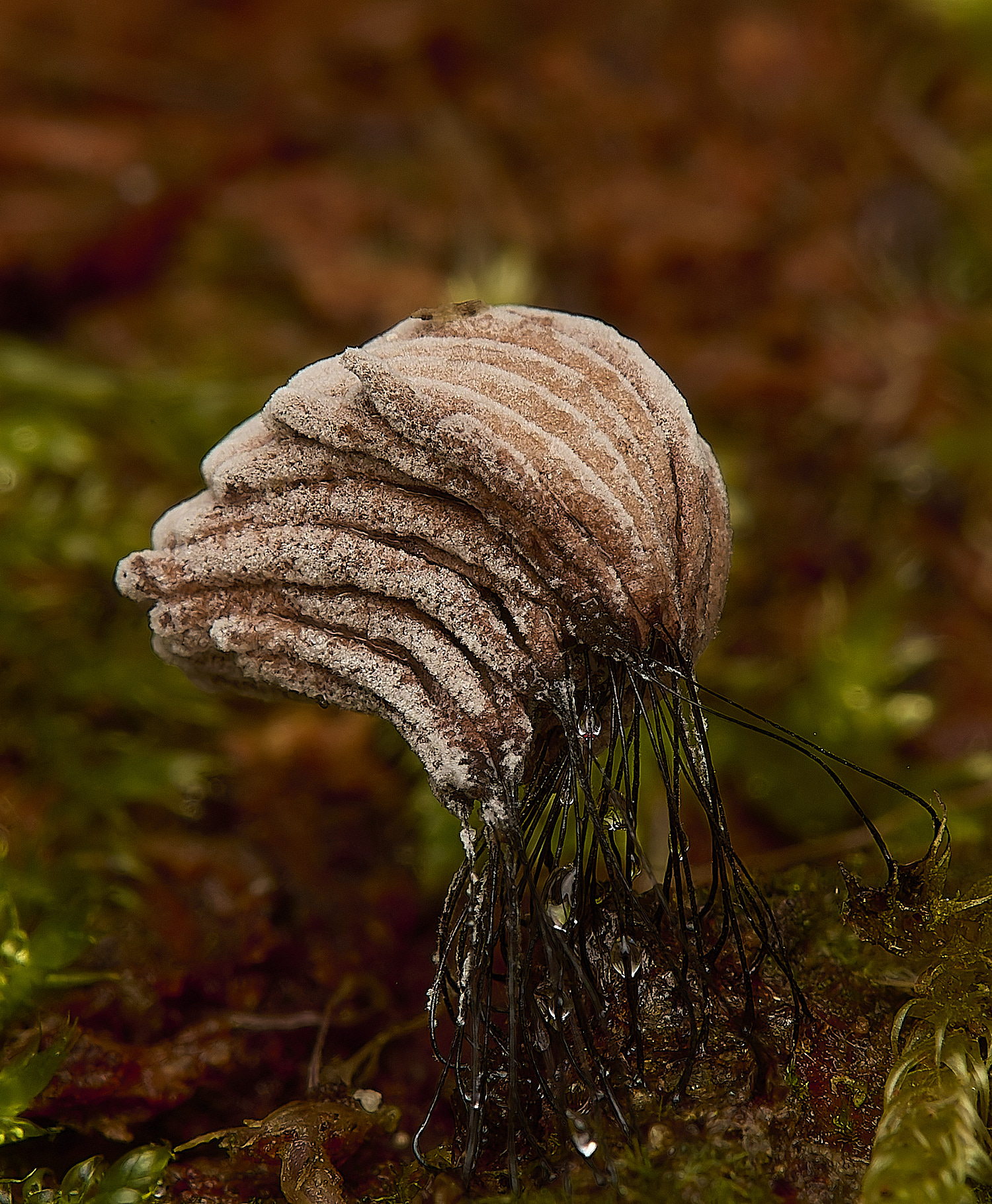
Stemonites fusca?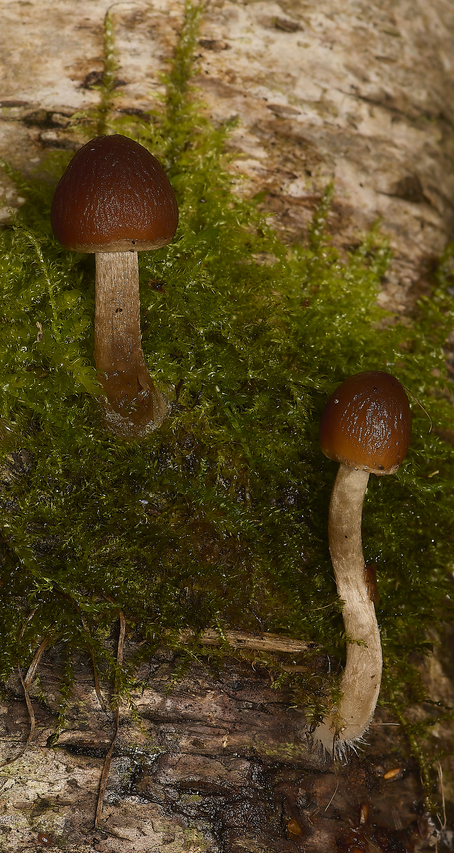
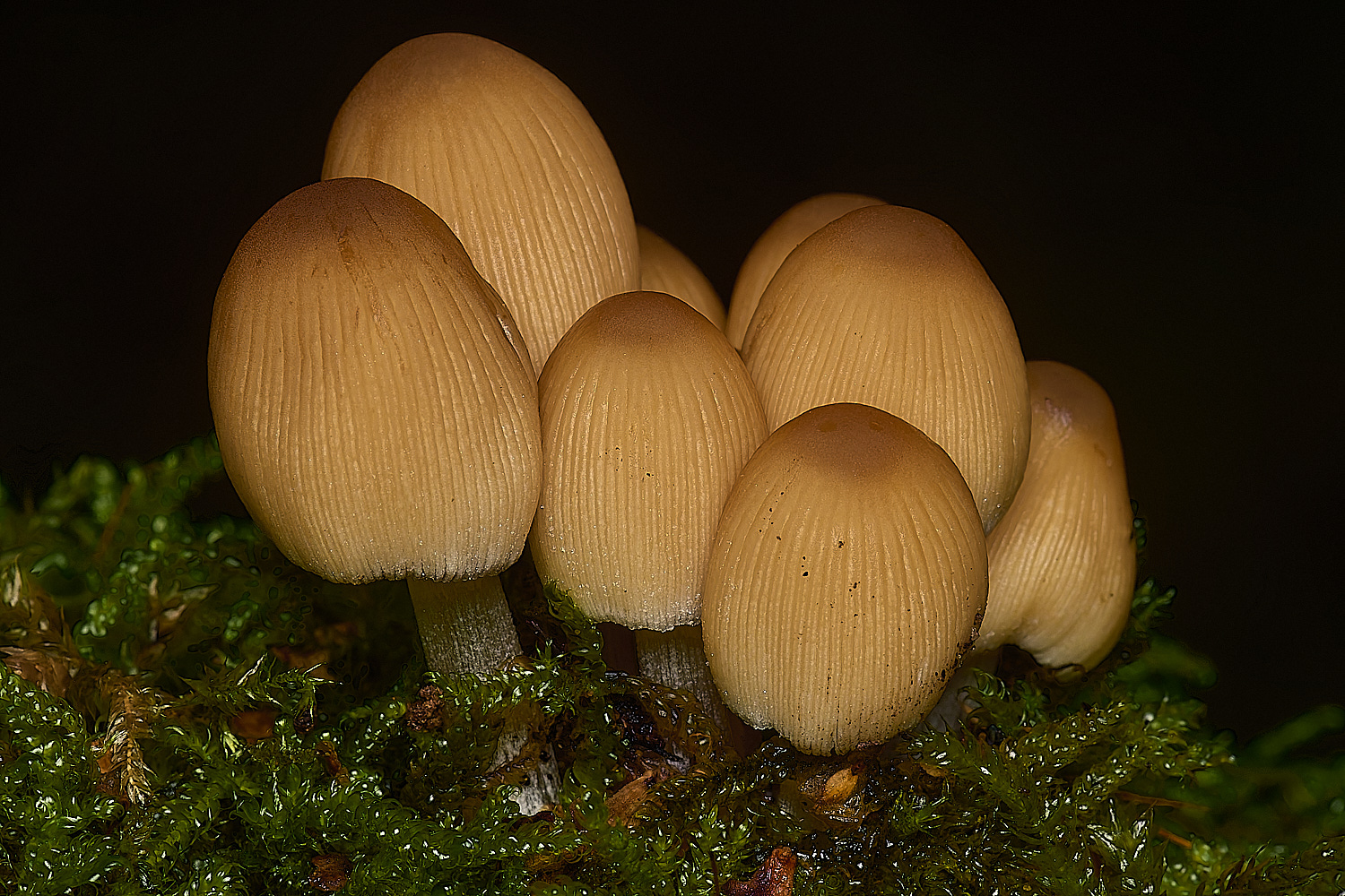
Stump Brittlestem (Psyatherella piluliformis)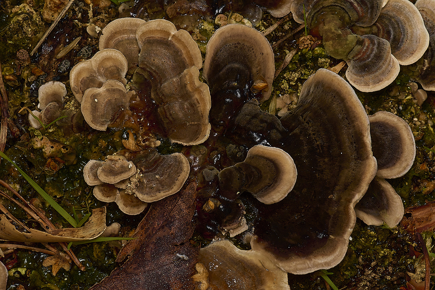
Turkey tail (Trametes versicolor)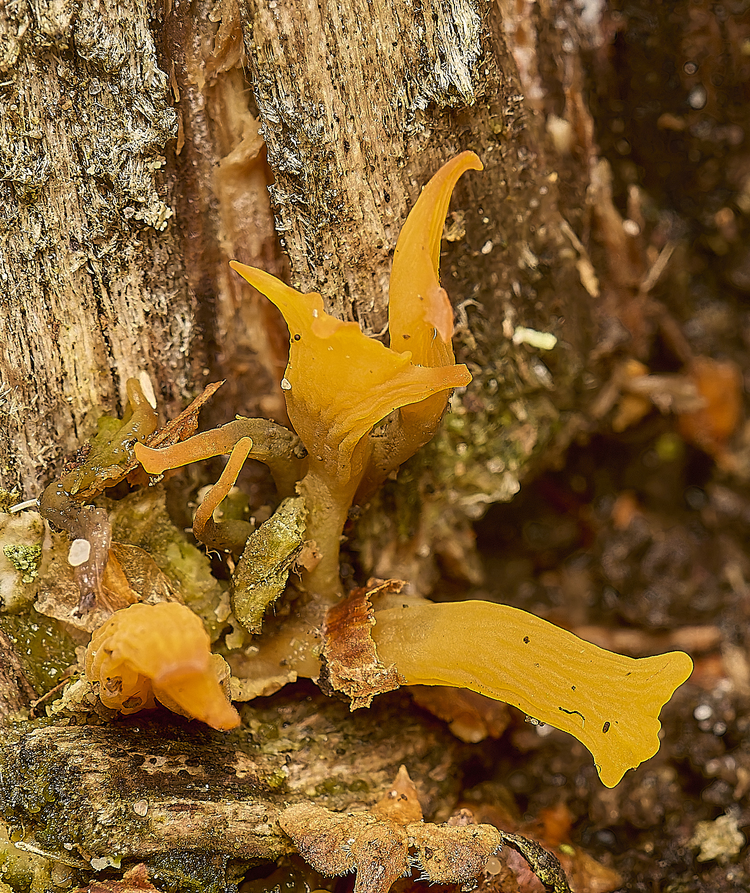
A stags horn on Oak (Quercus robur)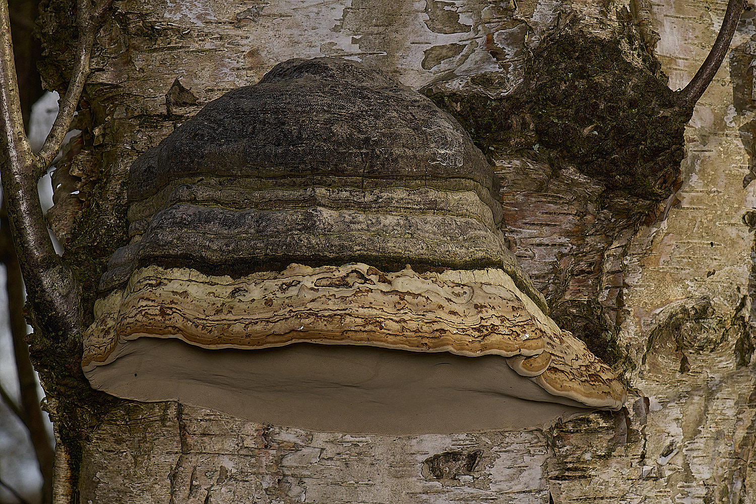
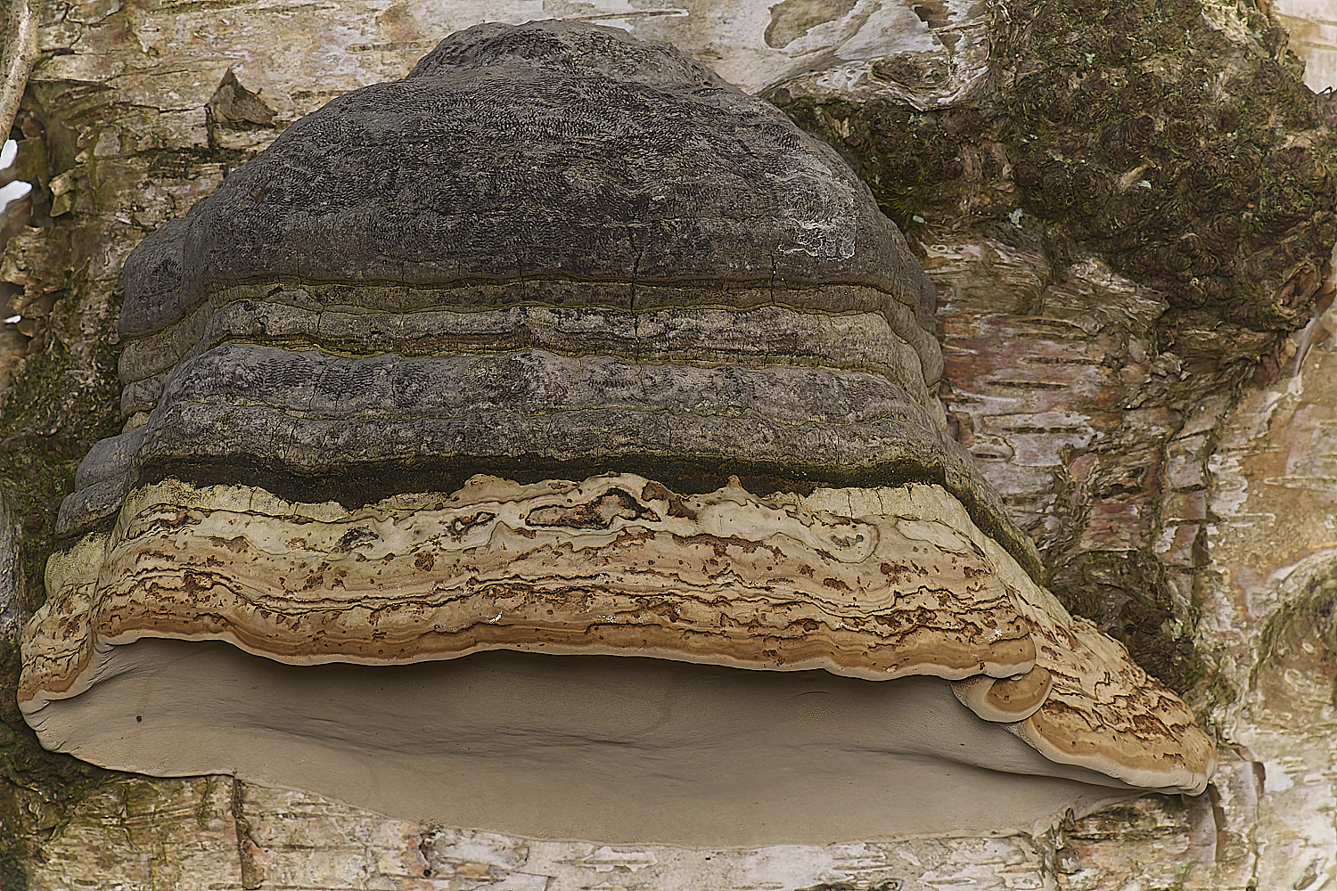
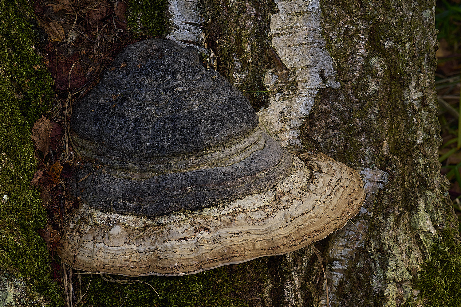
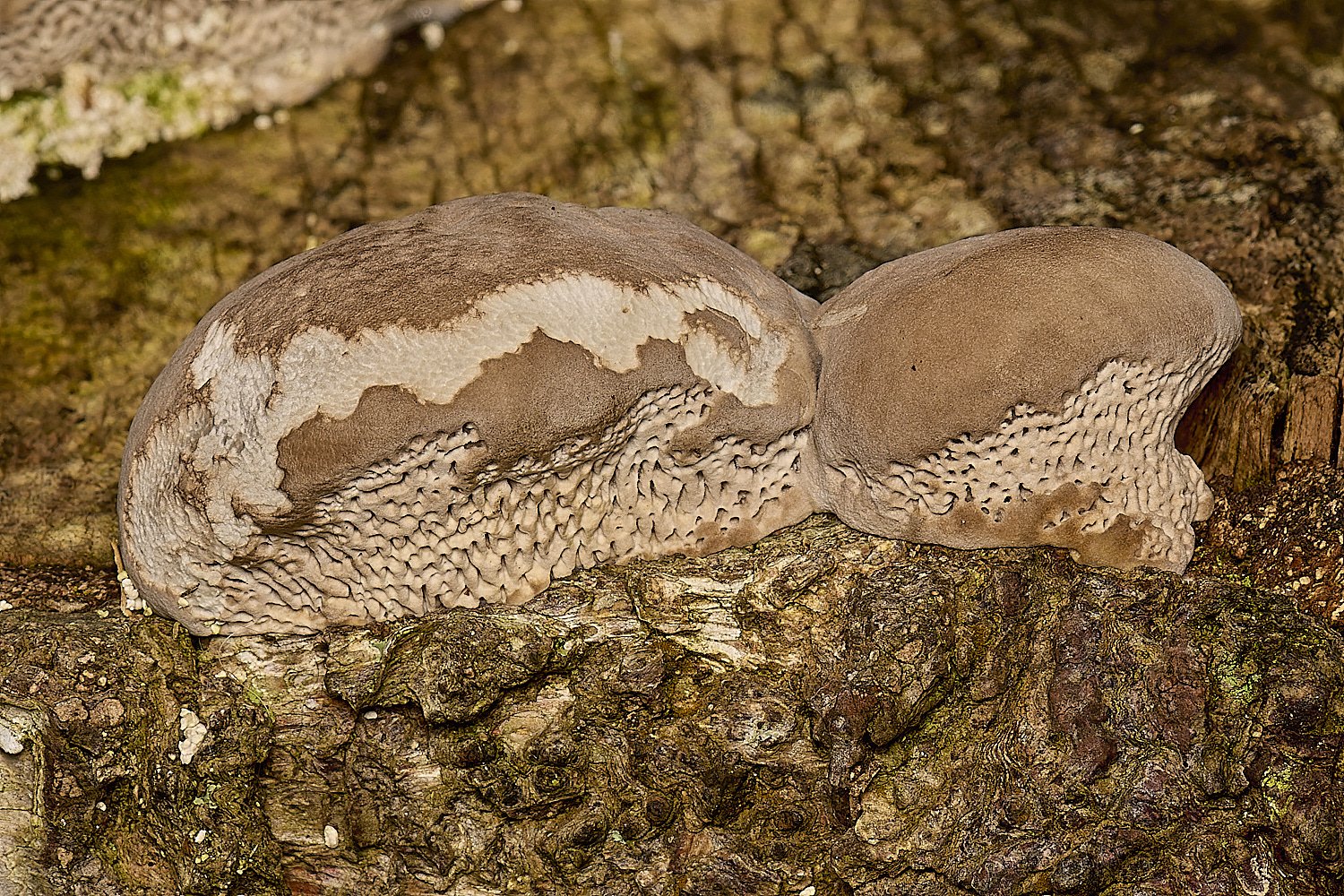
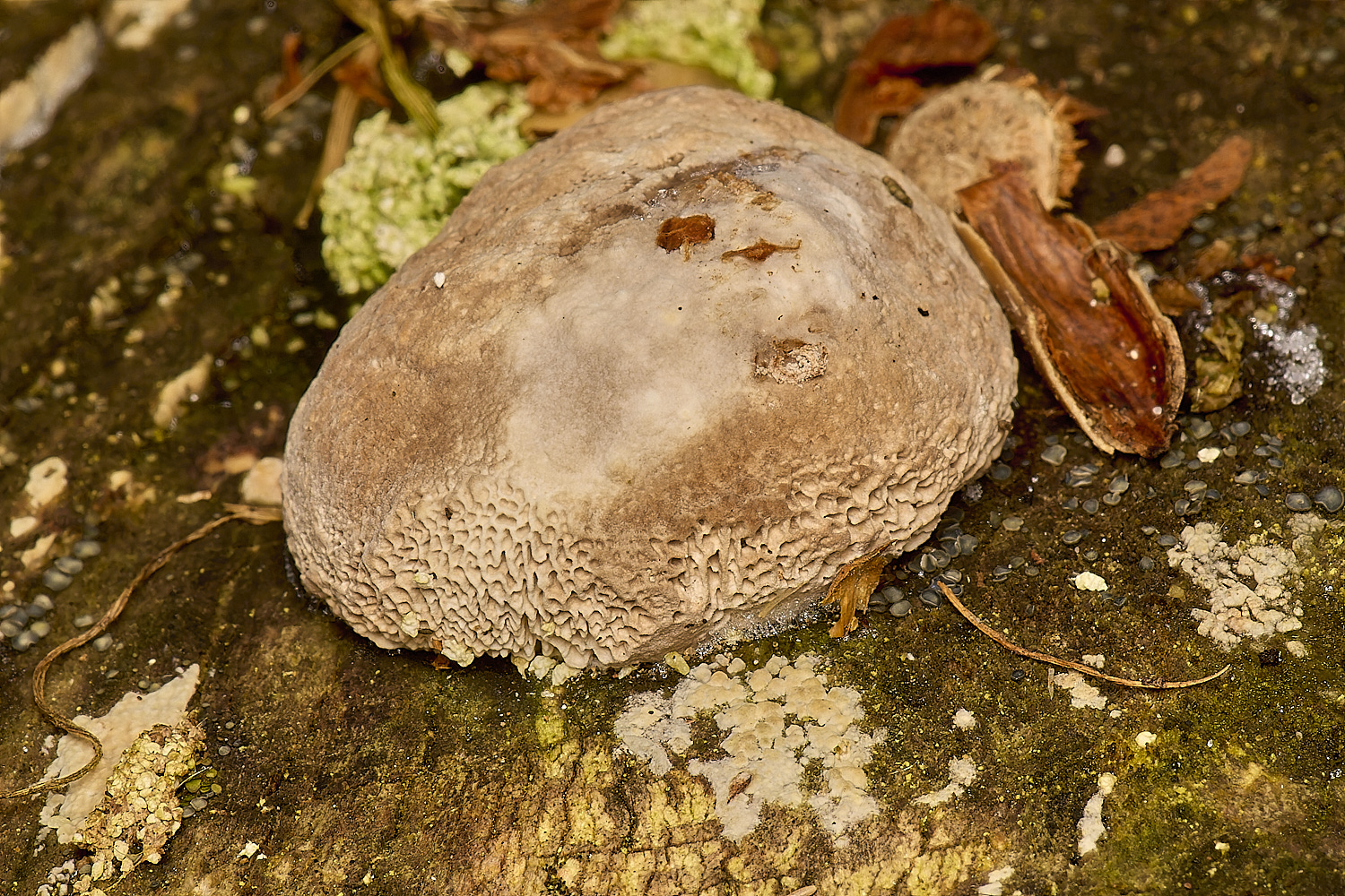
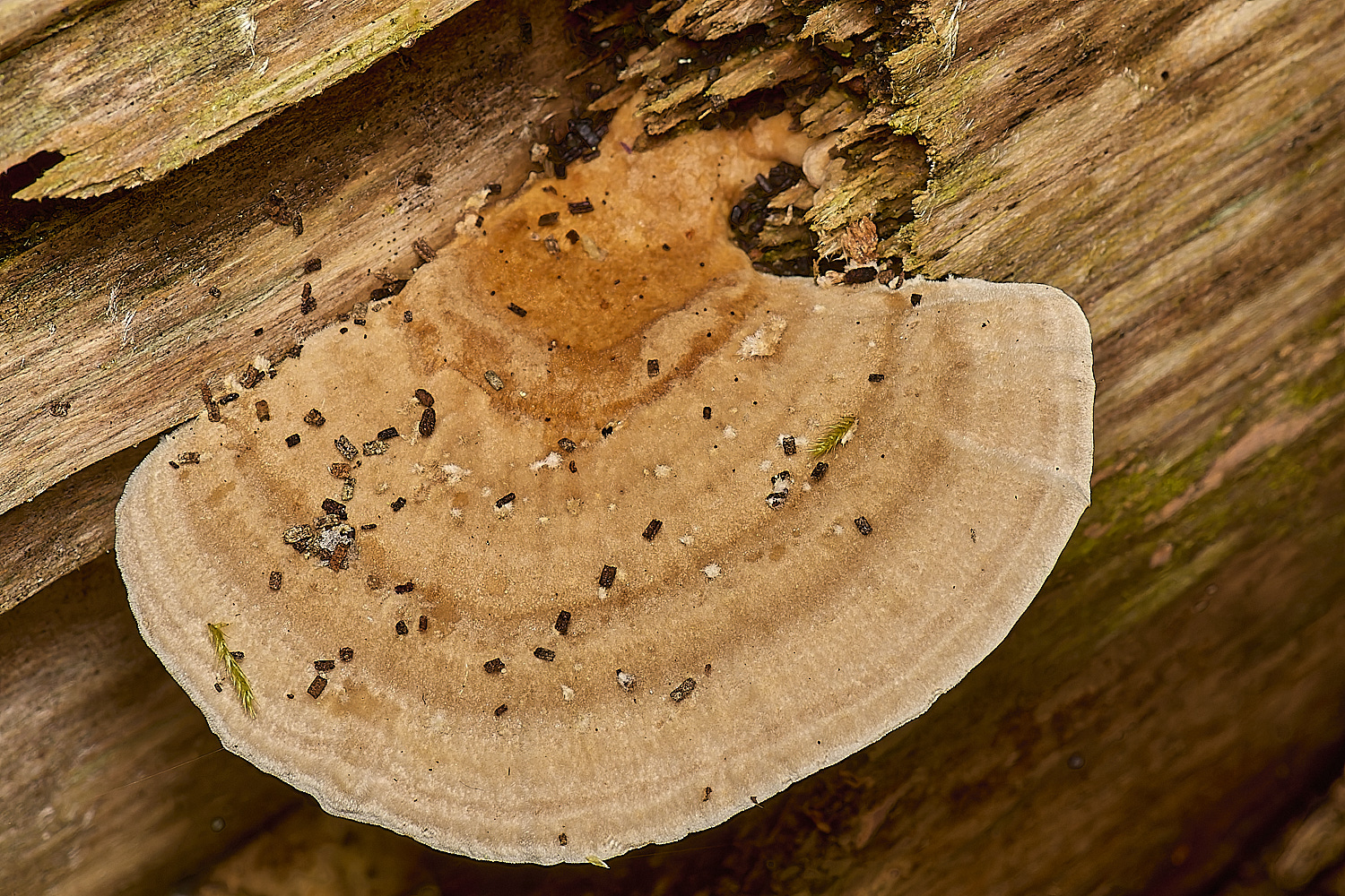
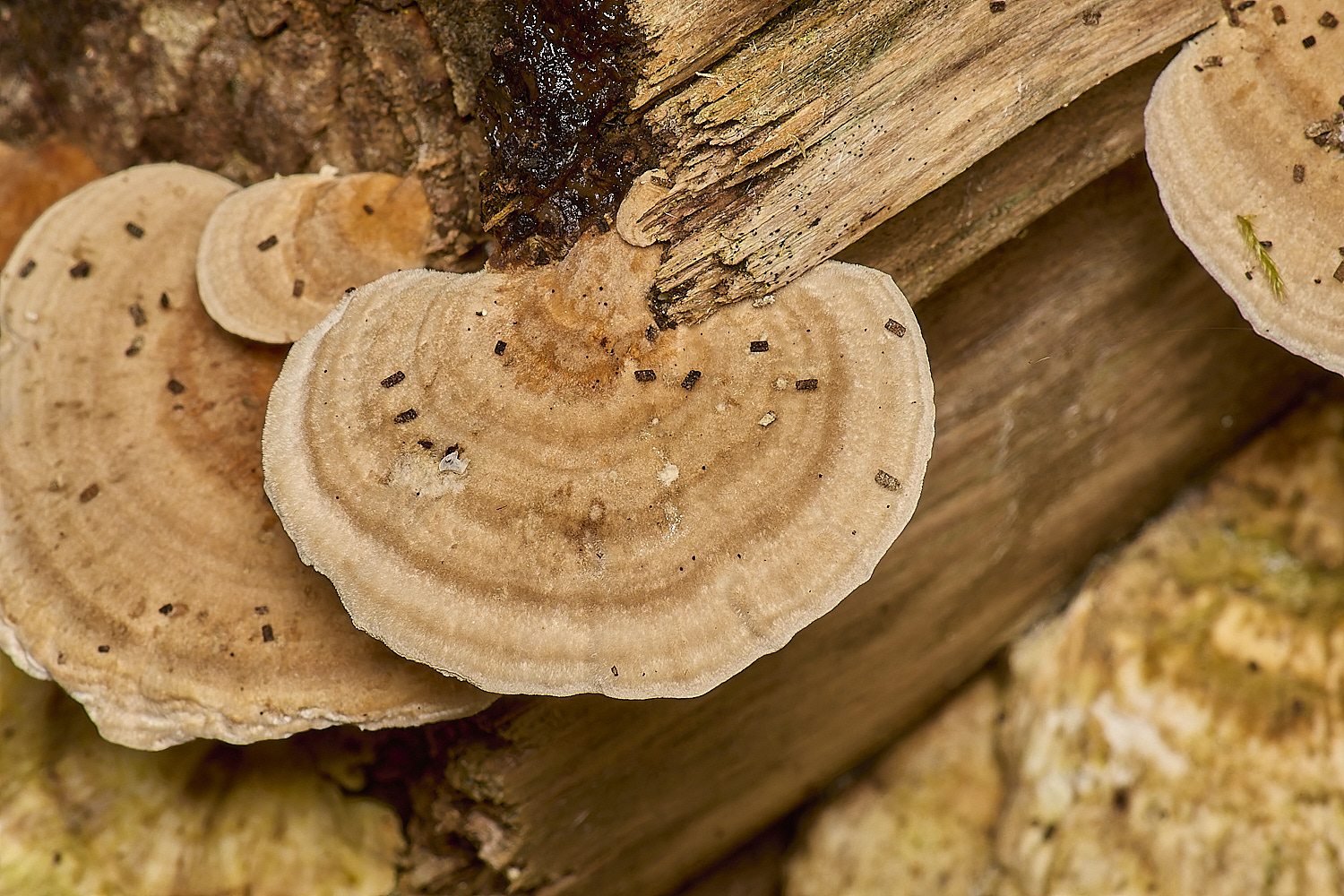
Hoof Fungus (Fomes fomentarius)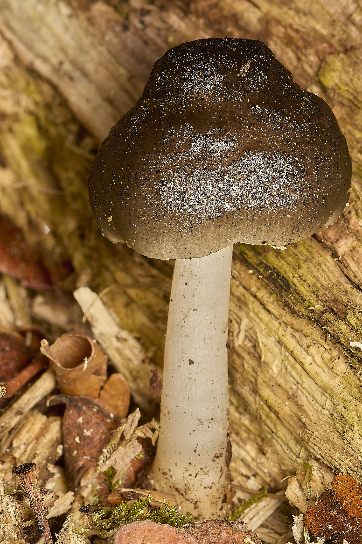
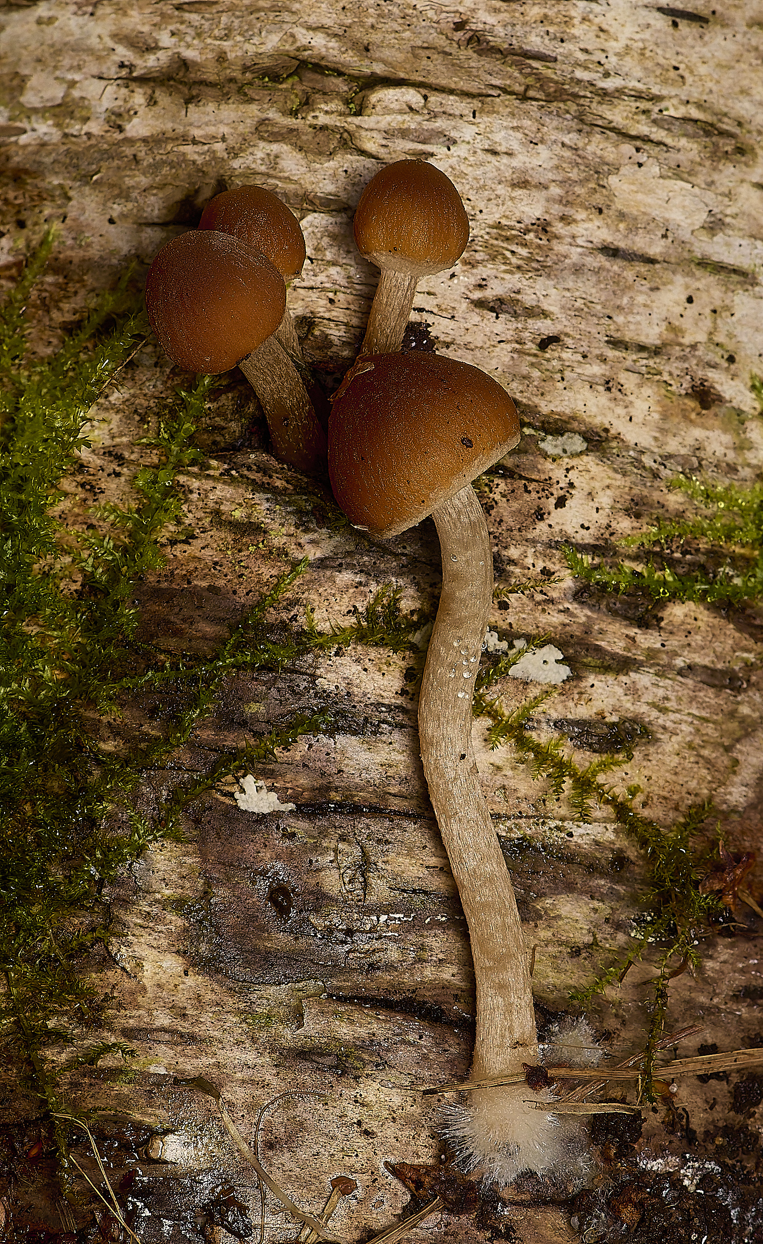
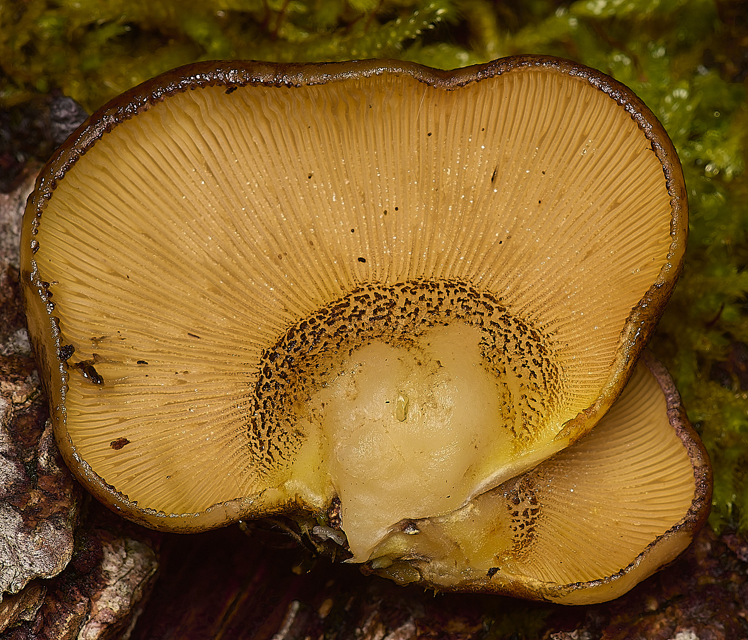
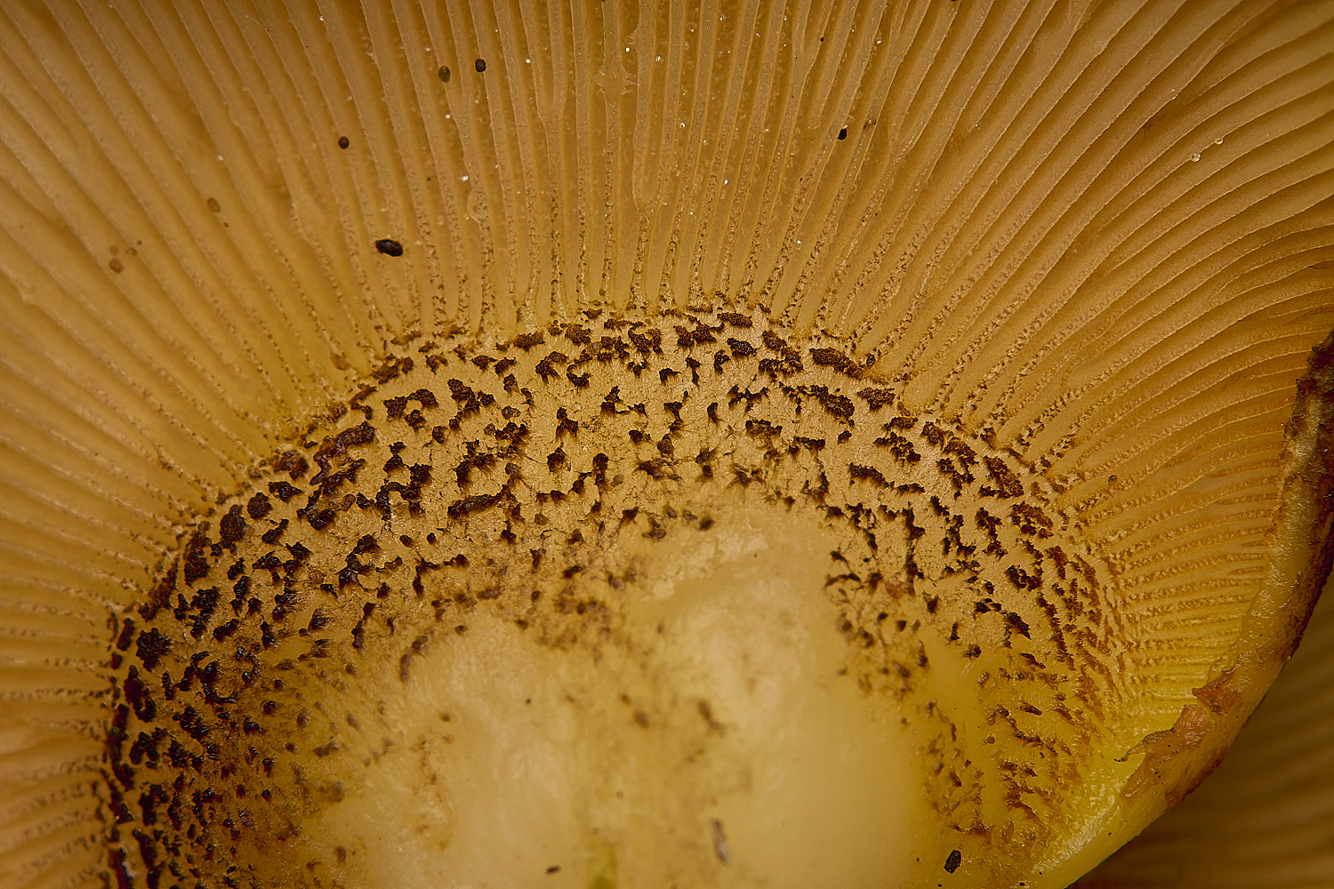
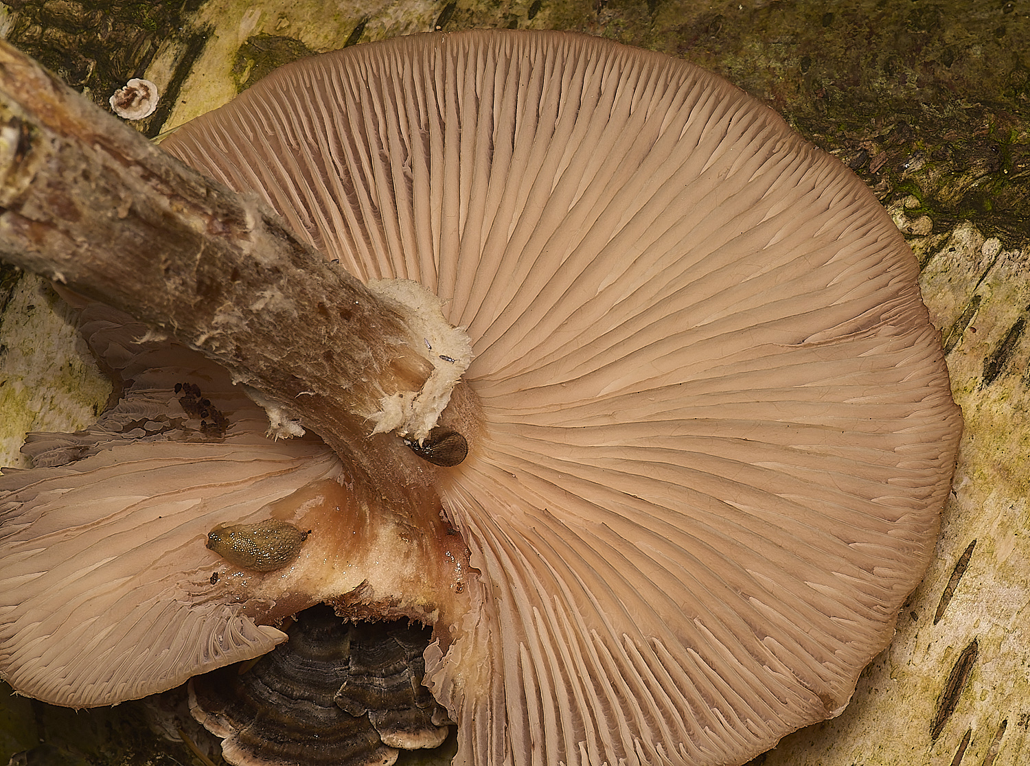
Deer Shield (Pluteus cervinus)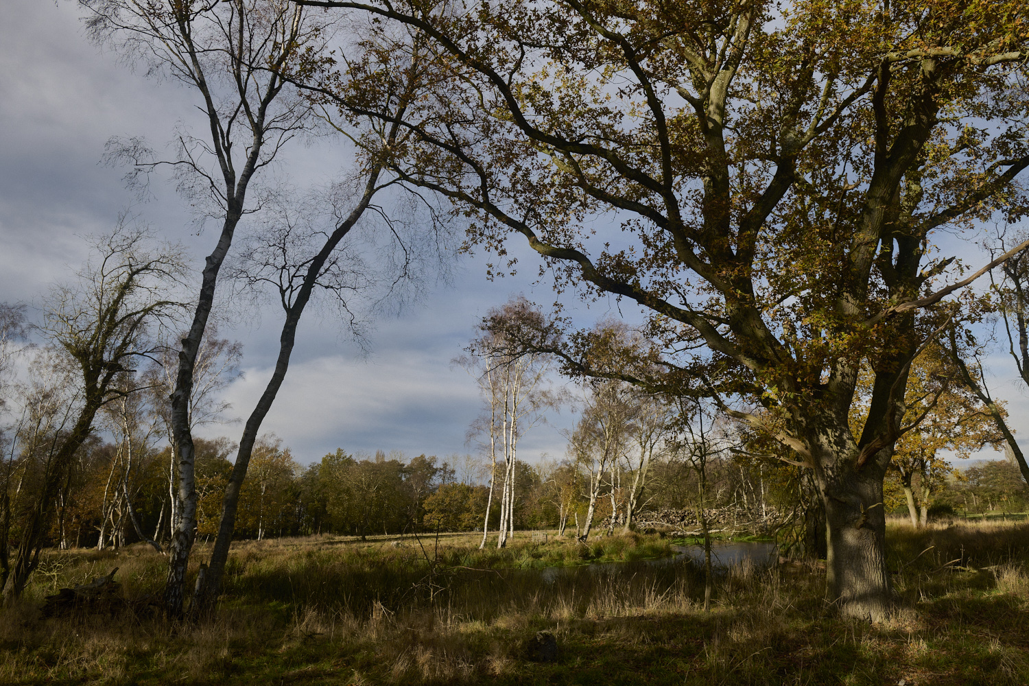
Hairy Curtain Crust (Stereum hirsutum)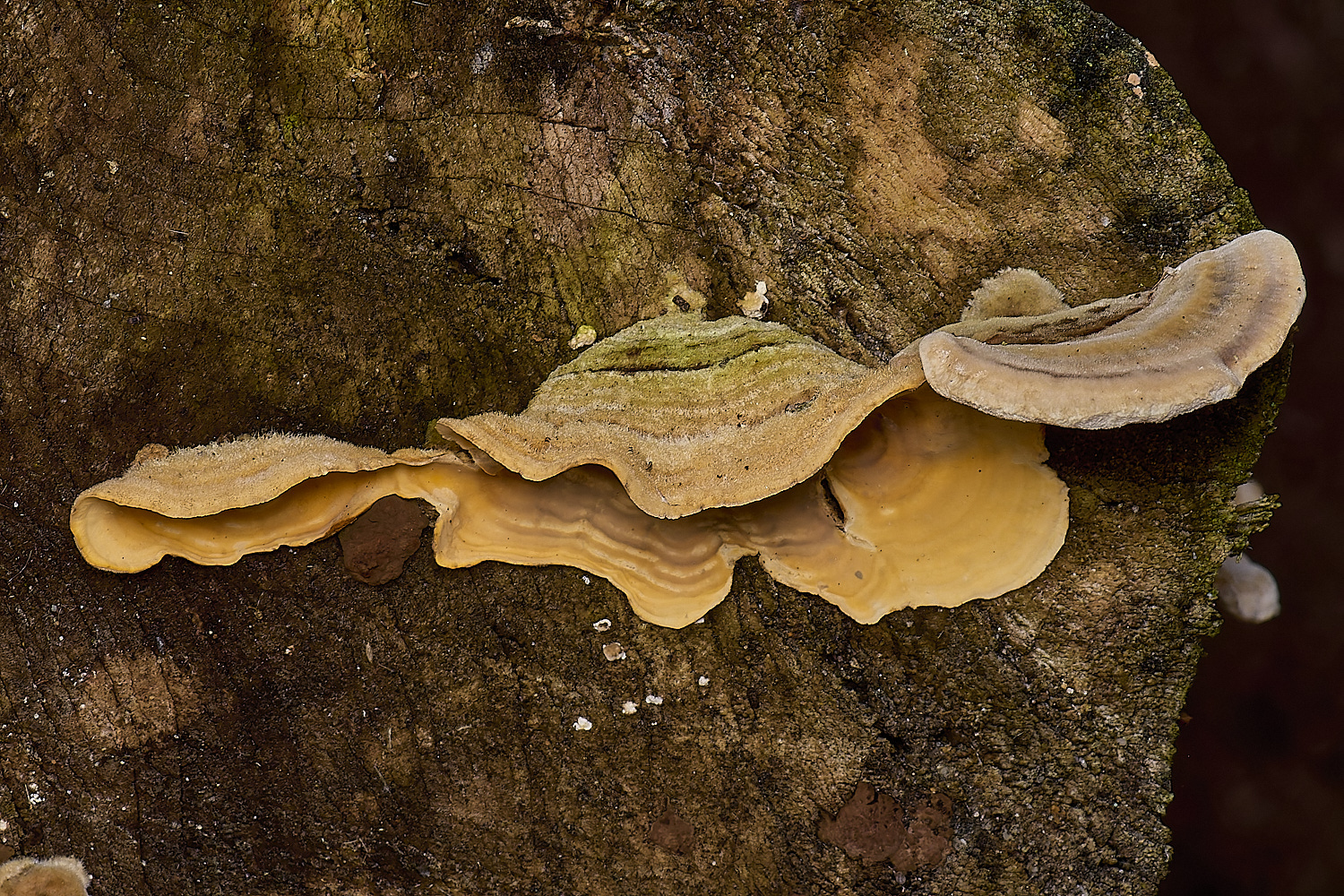
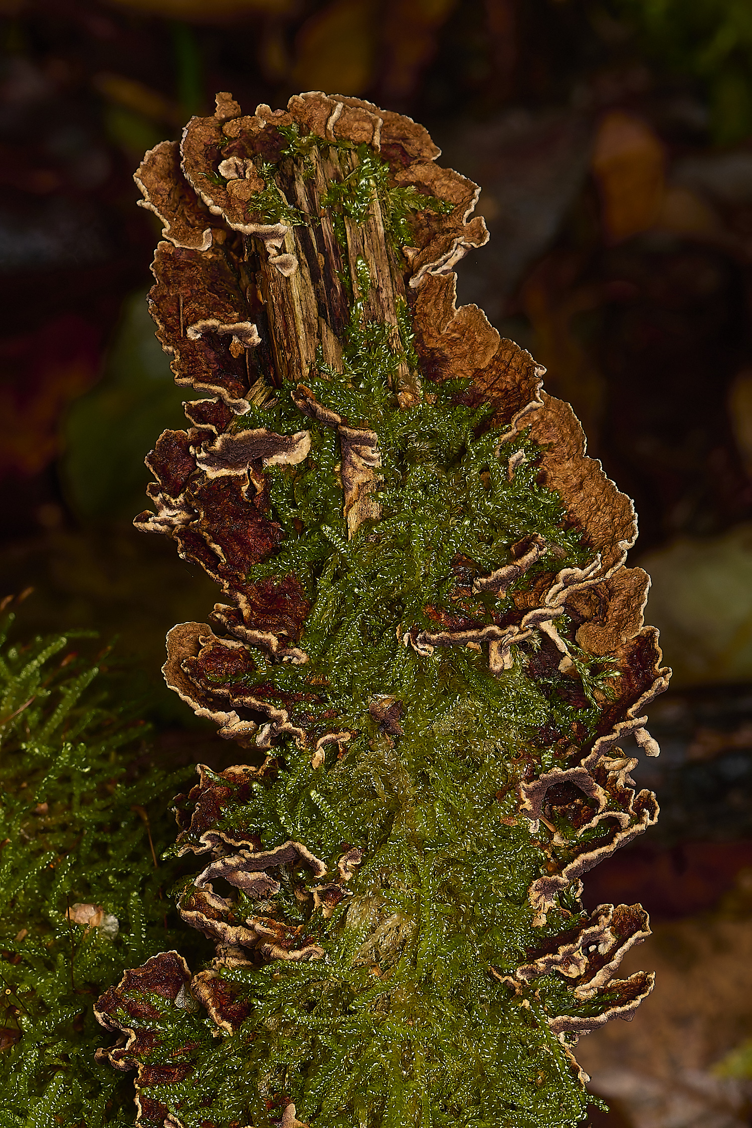
Turkey Tail (Trmaetes versicolor)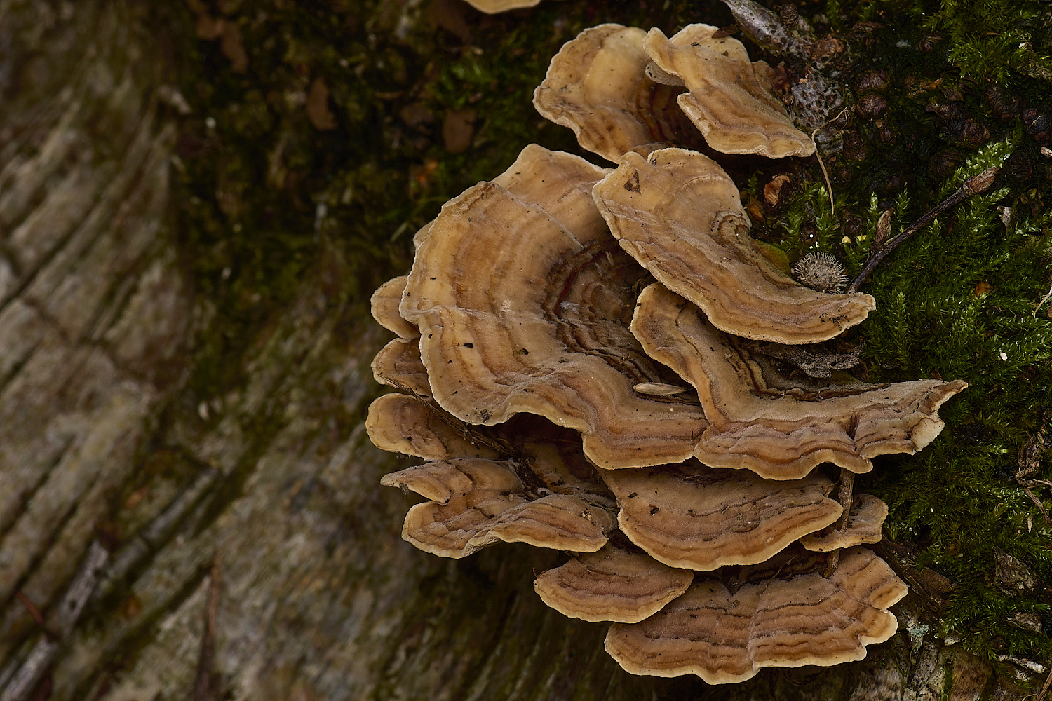
Yellowing Curtain Crust (Stereum submentosum)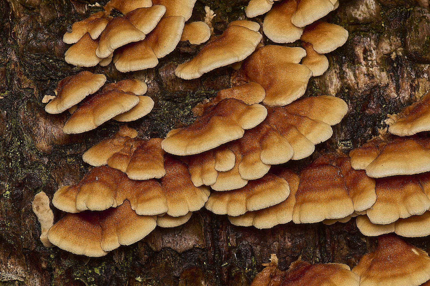
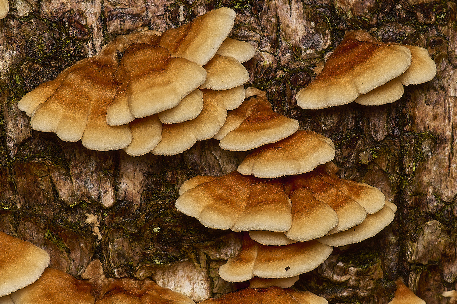
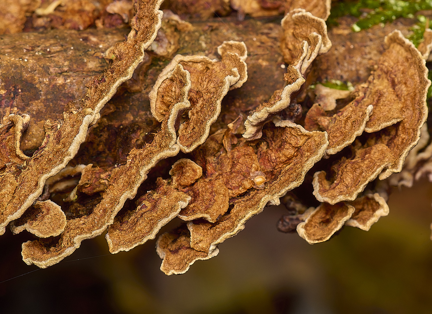
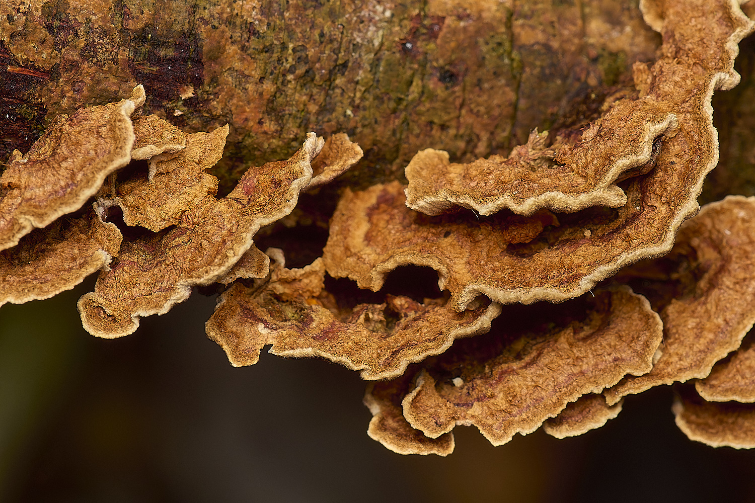
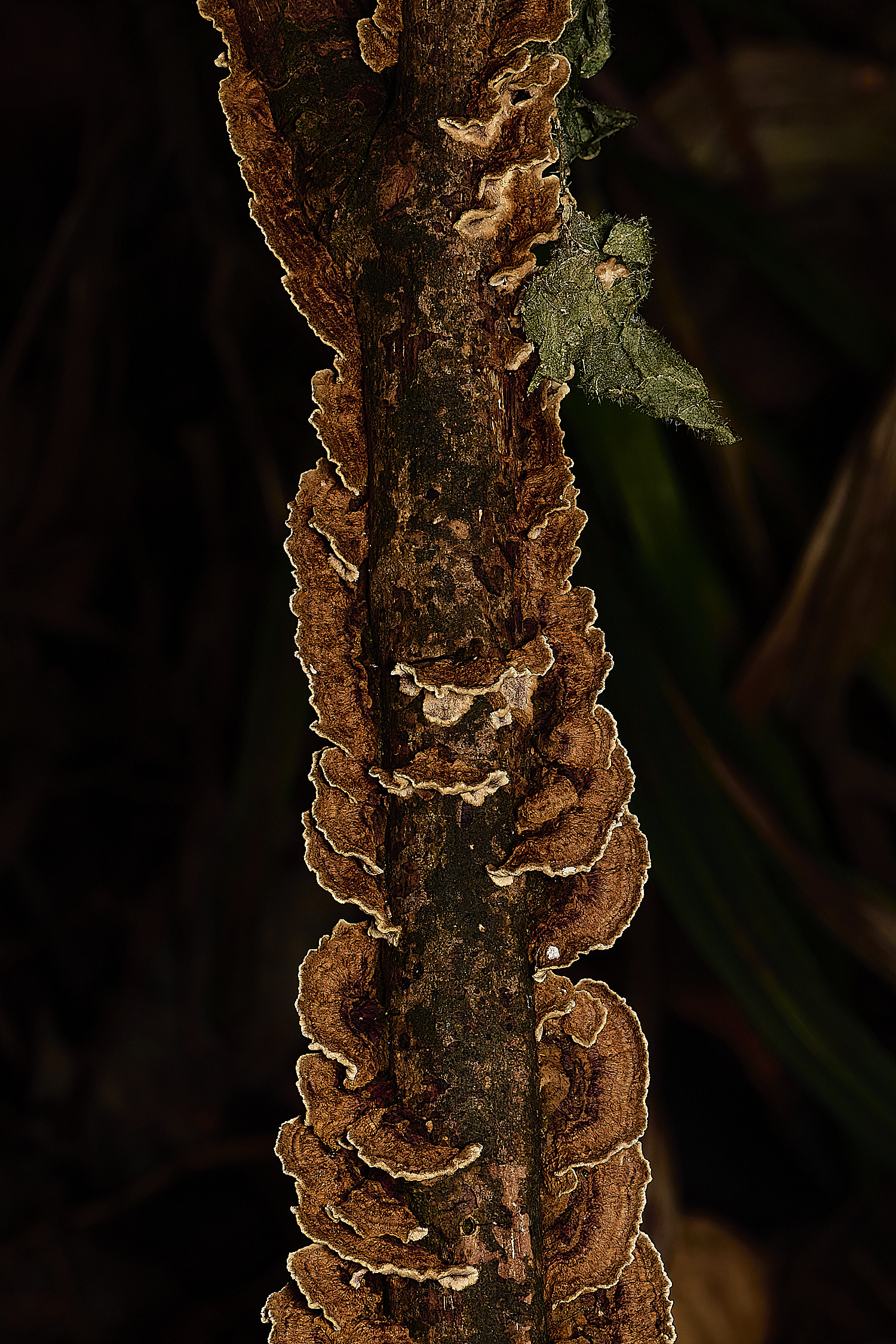
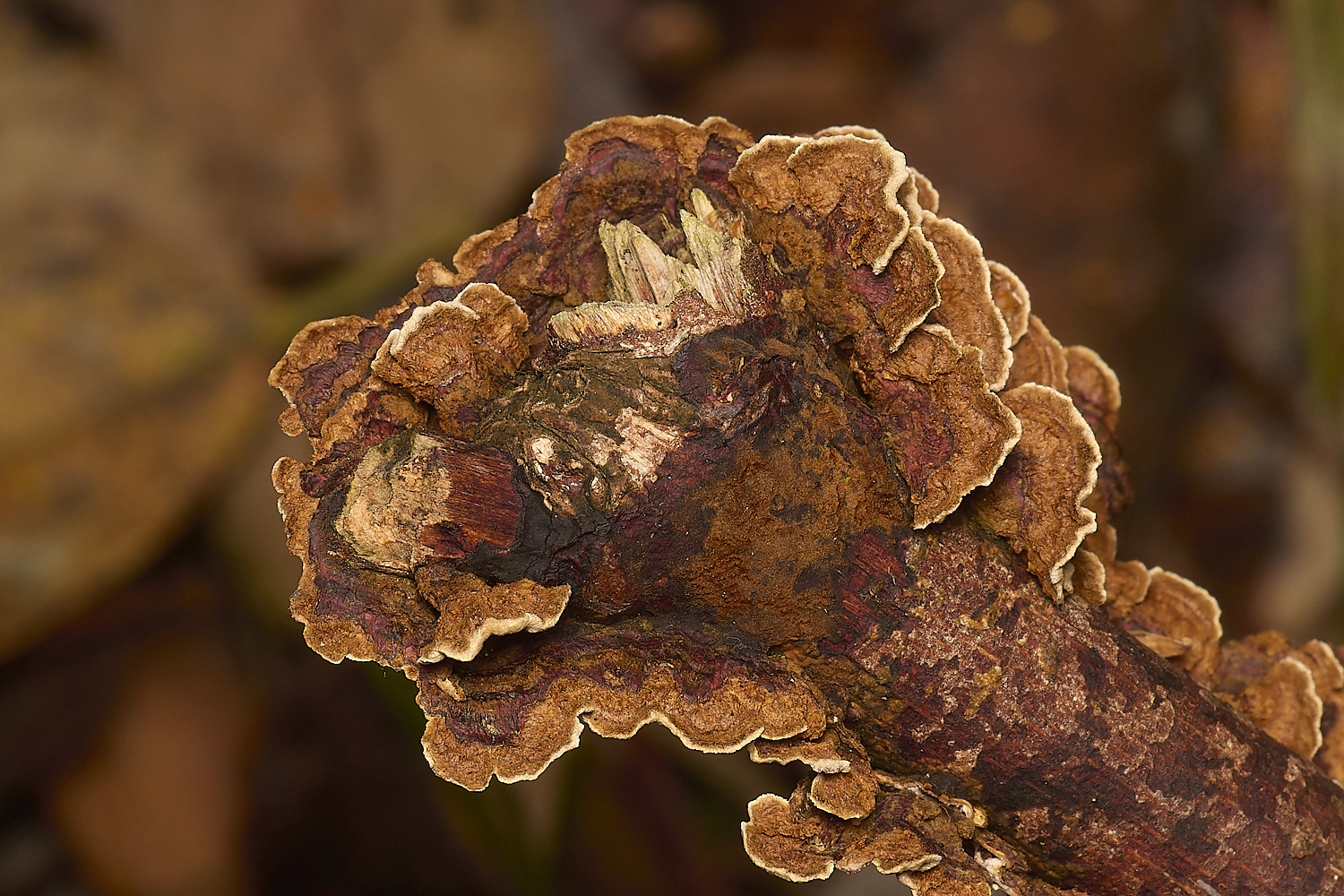
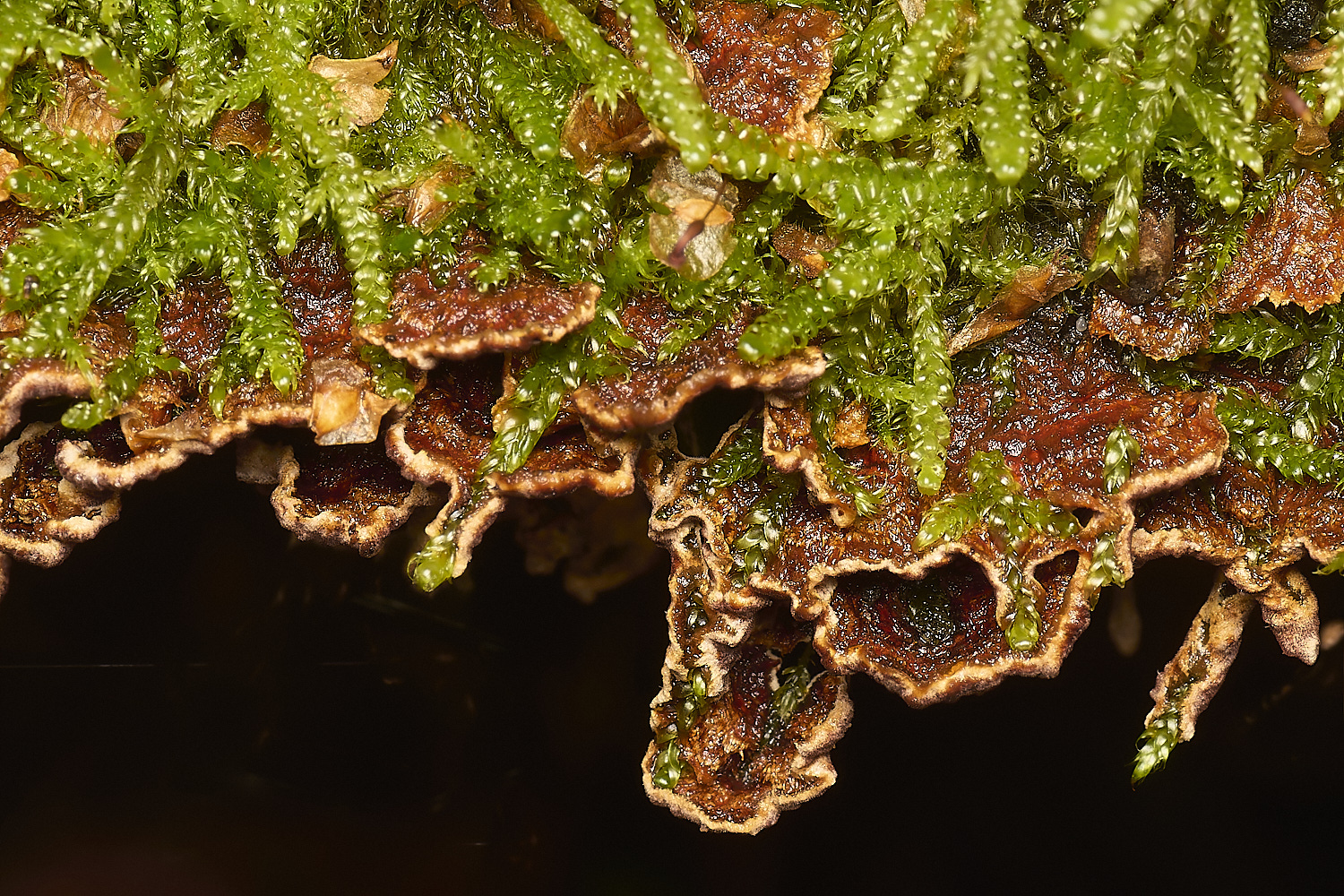
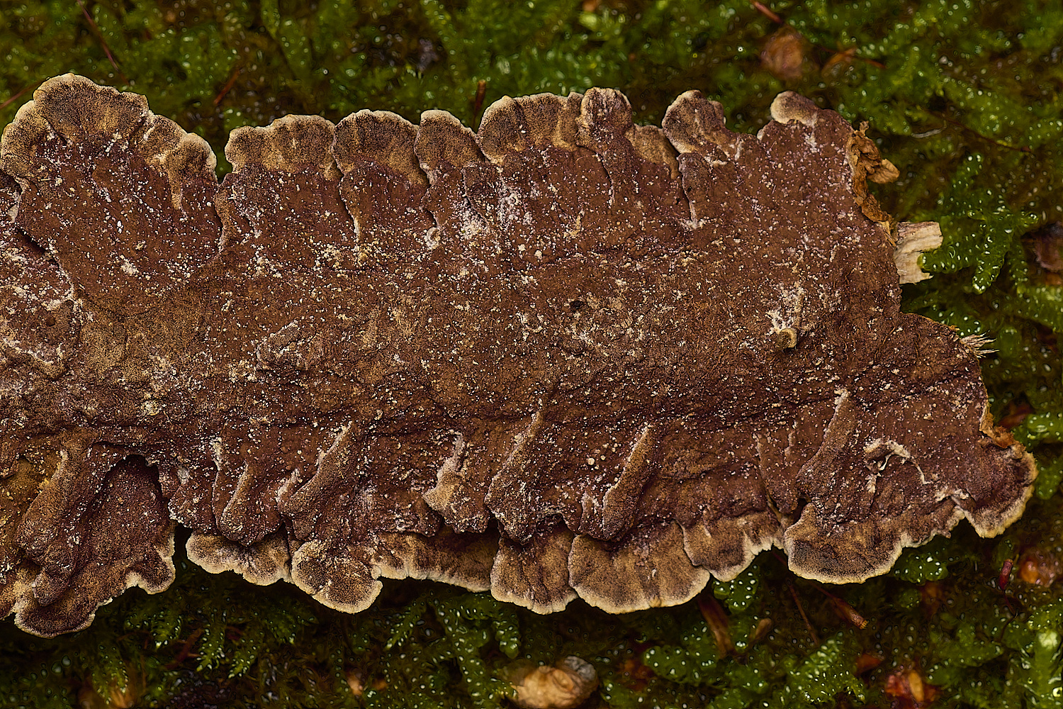
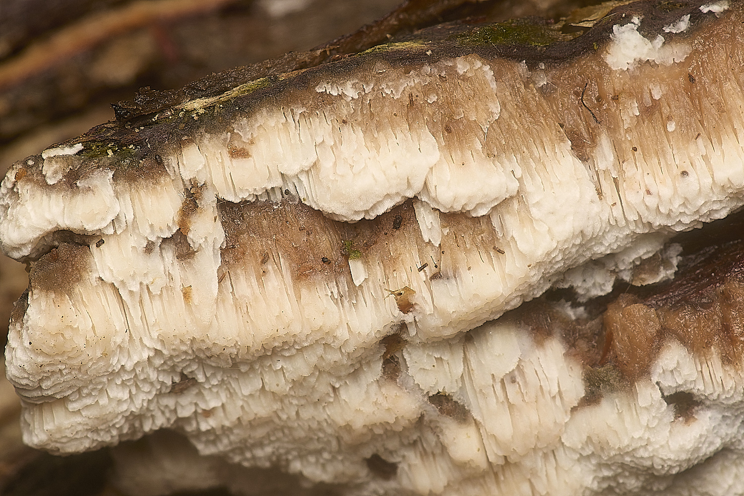
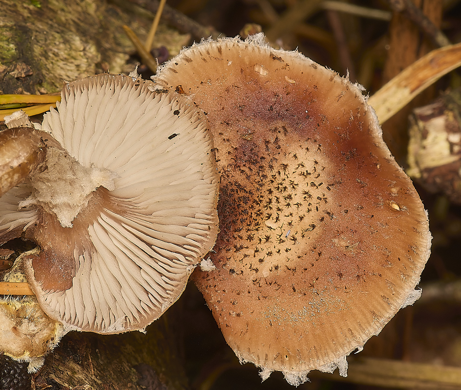
Crimp Gill (Plicatura crispa)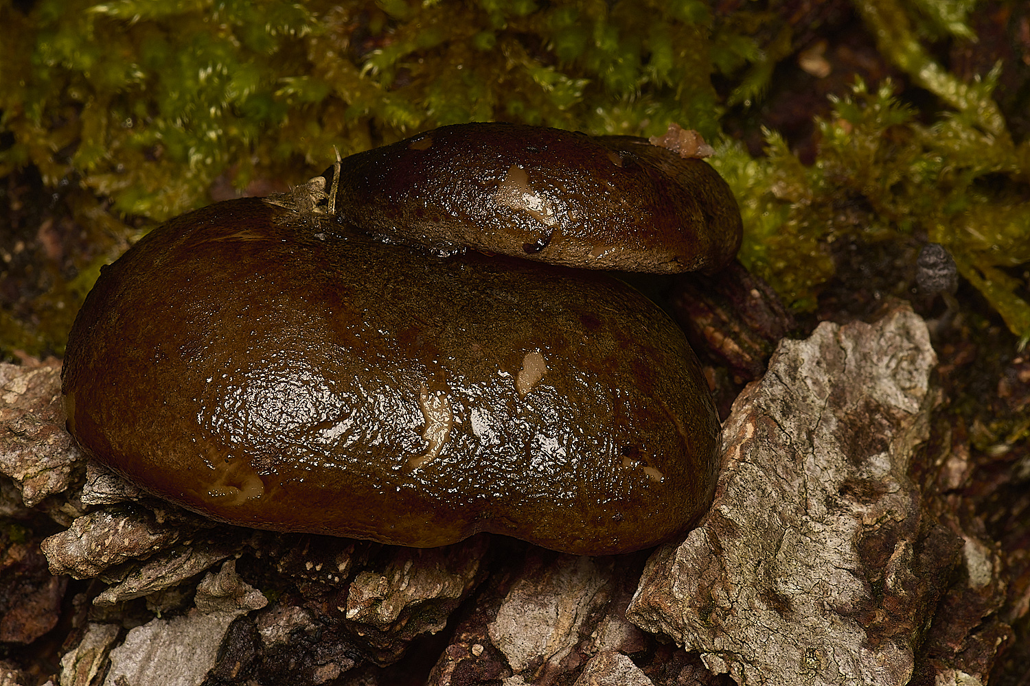
Olive Oysterling (Sarcomyxa serotina)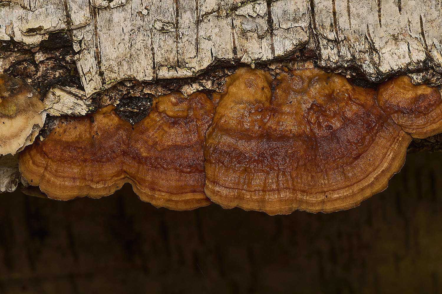
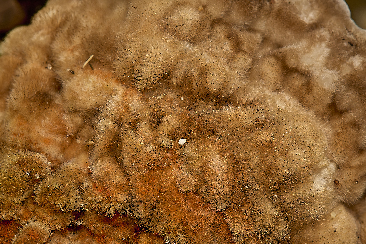
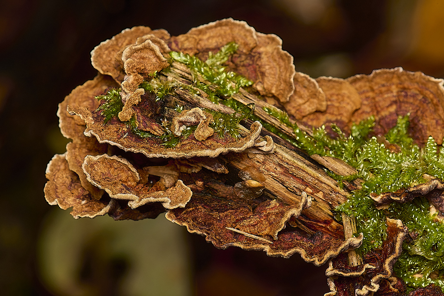
(Trametes pubescens)
Much paler (Although the top image belies that idea) than T versicolor and slightly hairy on the upper surface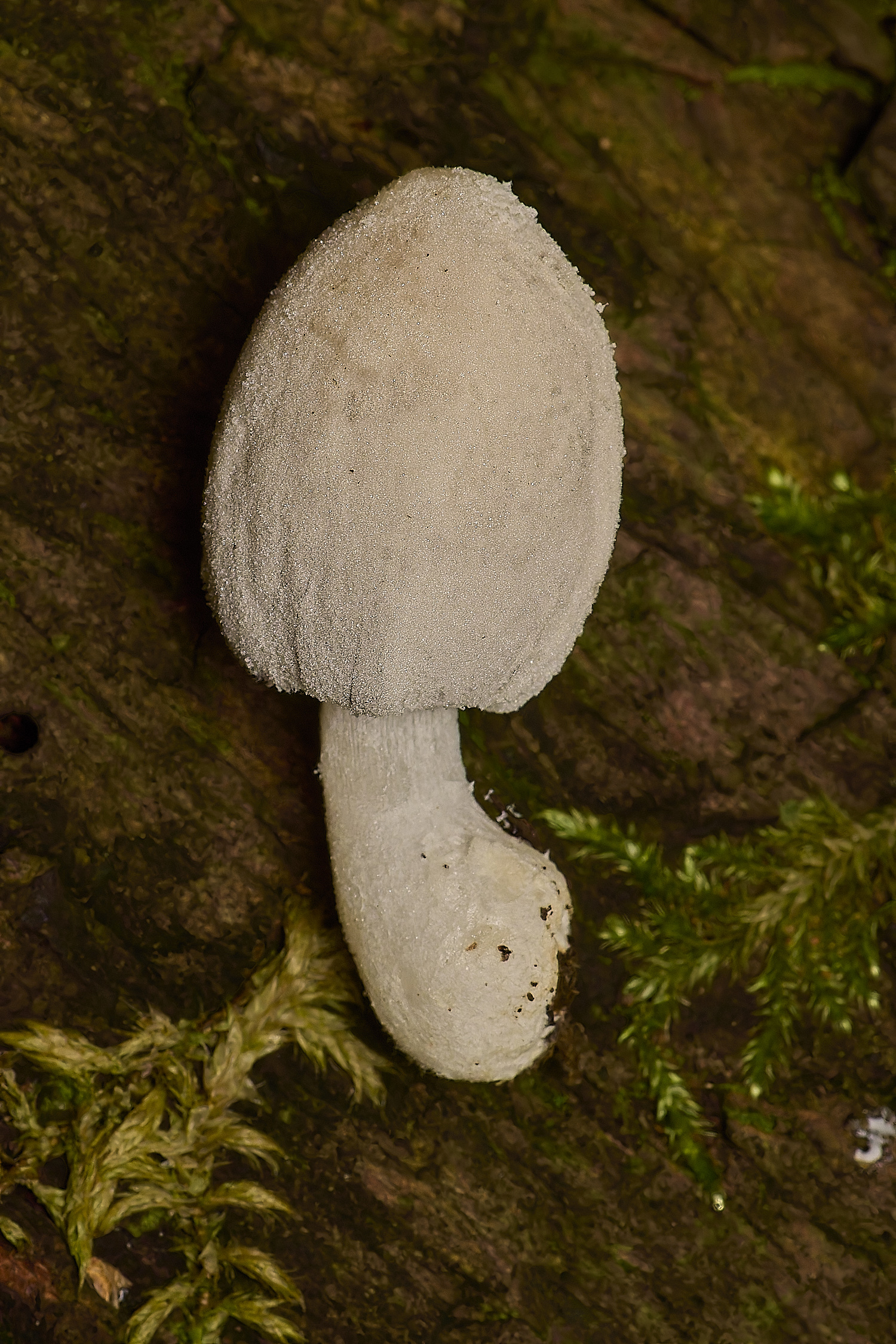
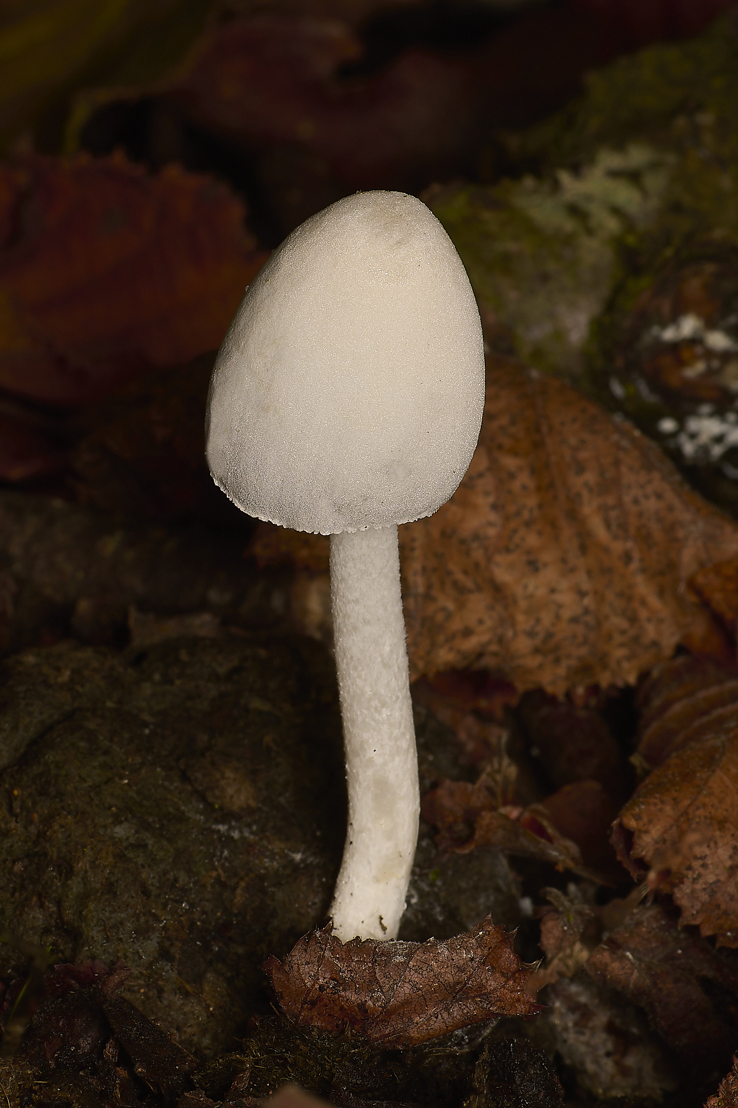
Snowy Inkcap (Coprinopsis nivea)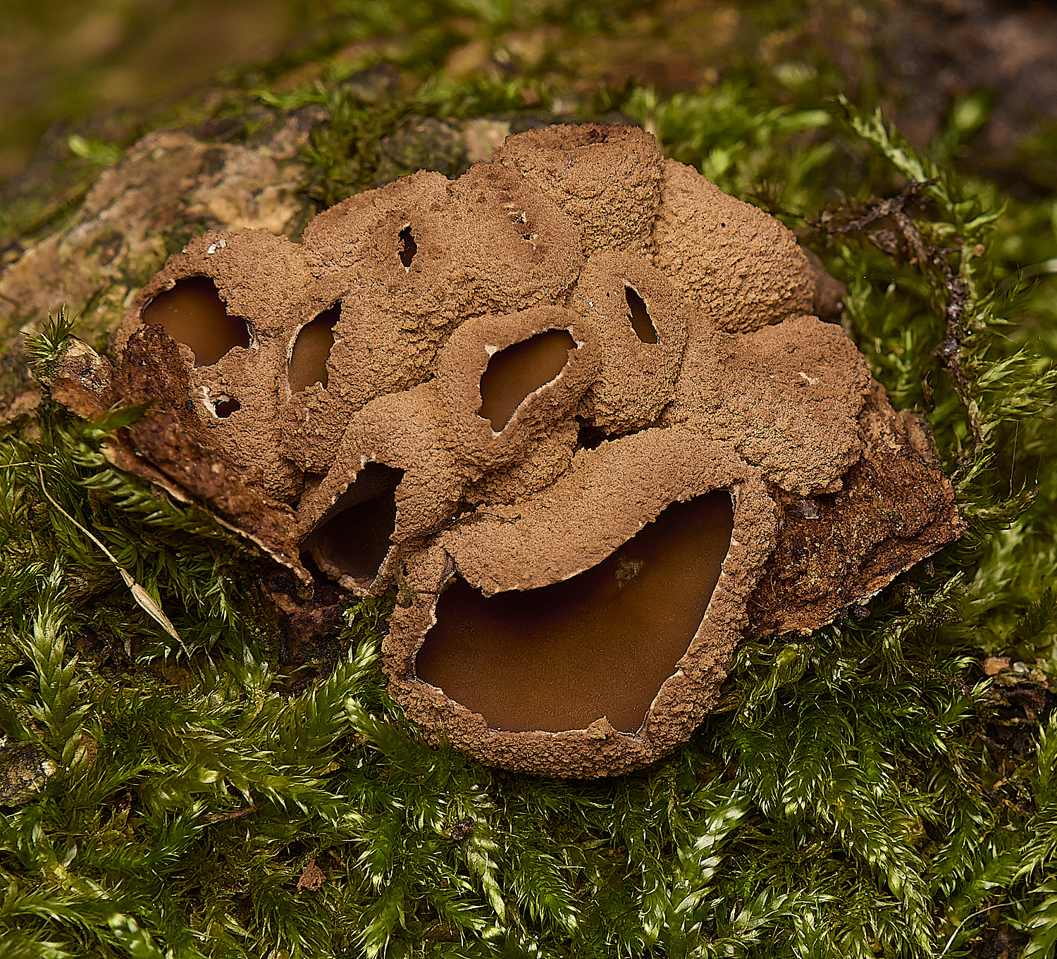
Spring Hazel Cup (Encoelia furfuracea)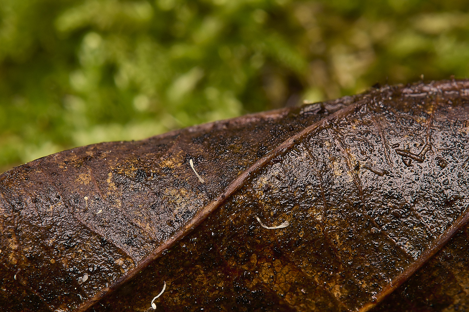
Tiny Typhula setipes on an Alder (Alnus glutinosa) leaf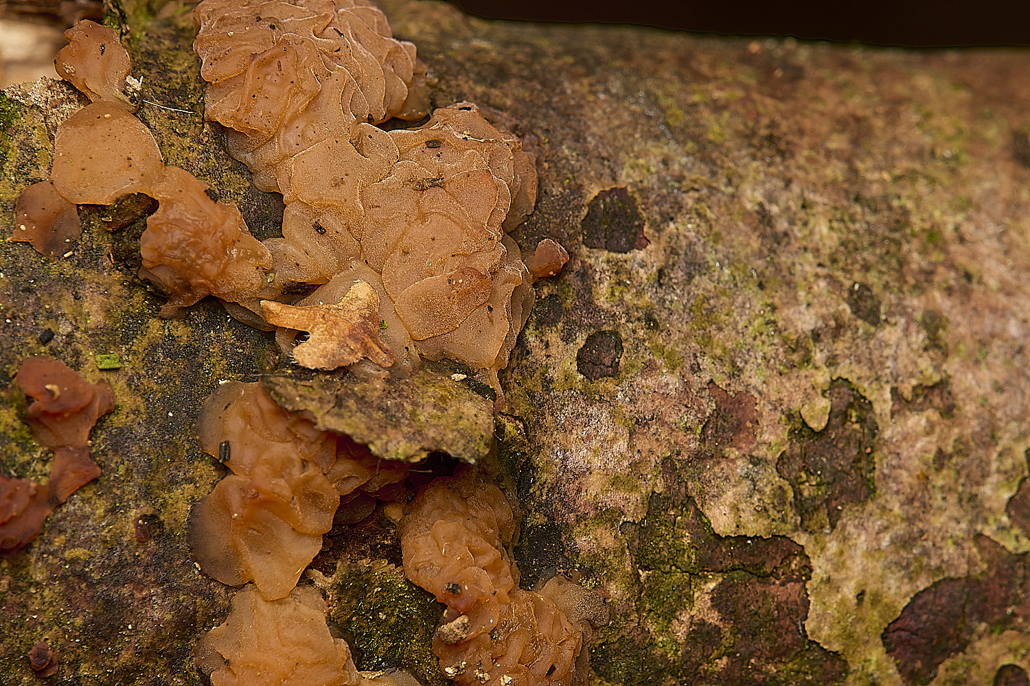
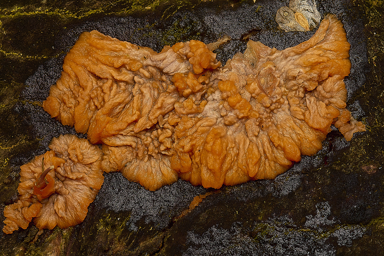
Leafy Brain (Phaeotremella frondosa) on Hazel (Corylus avellana)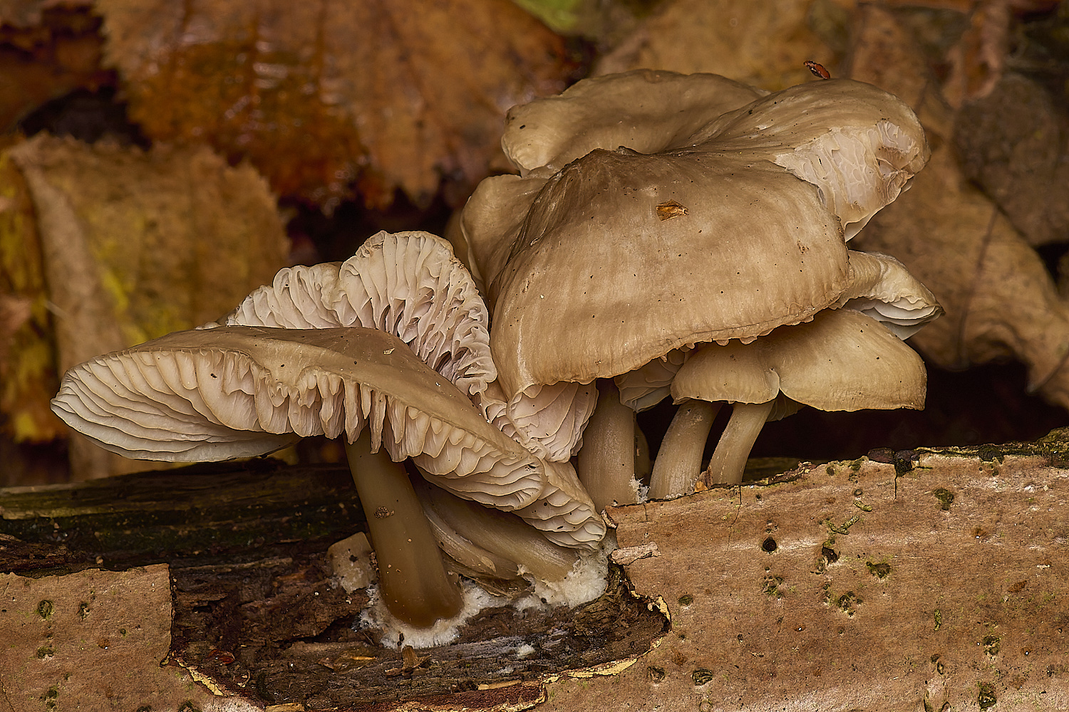
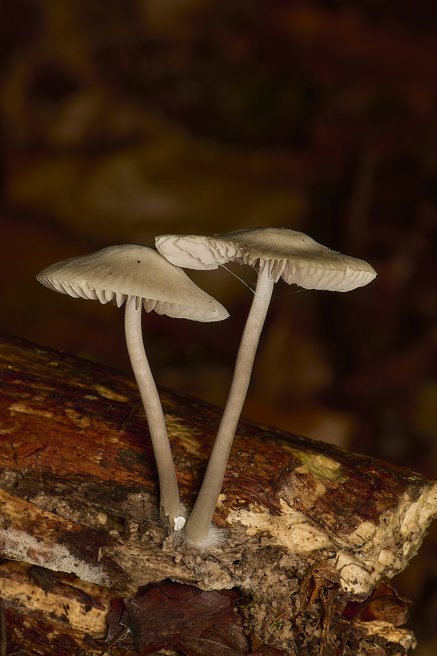
Common Bonnet (Mycena galericulata)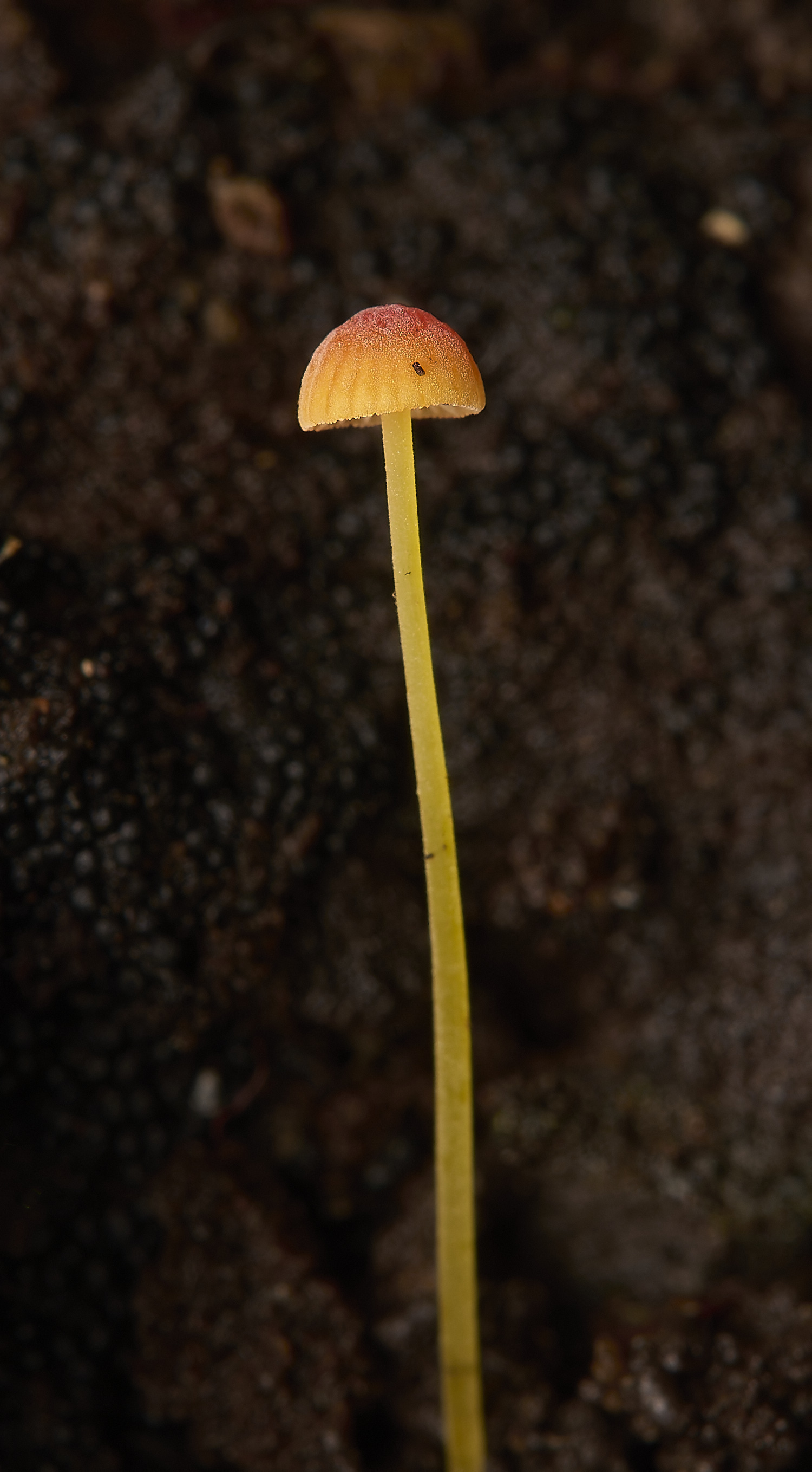
Orange Bonnet (Mycena acicula)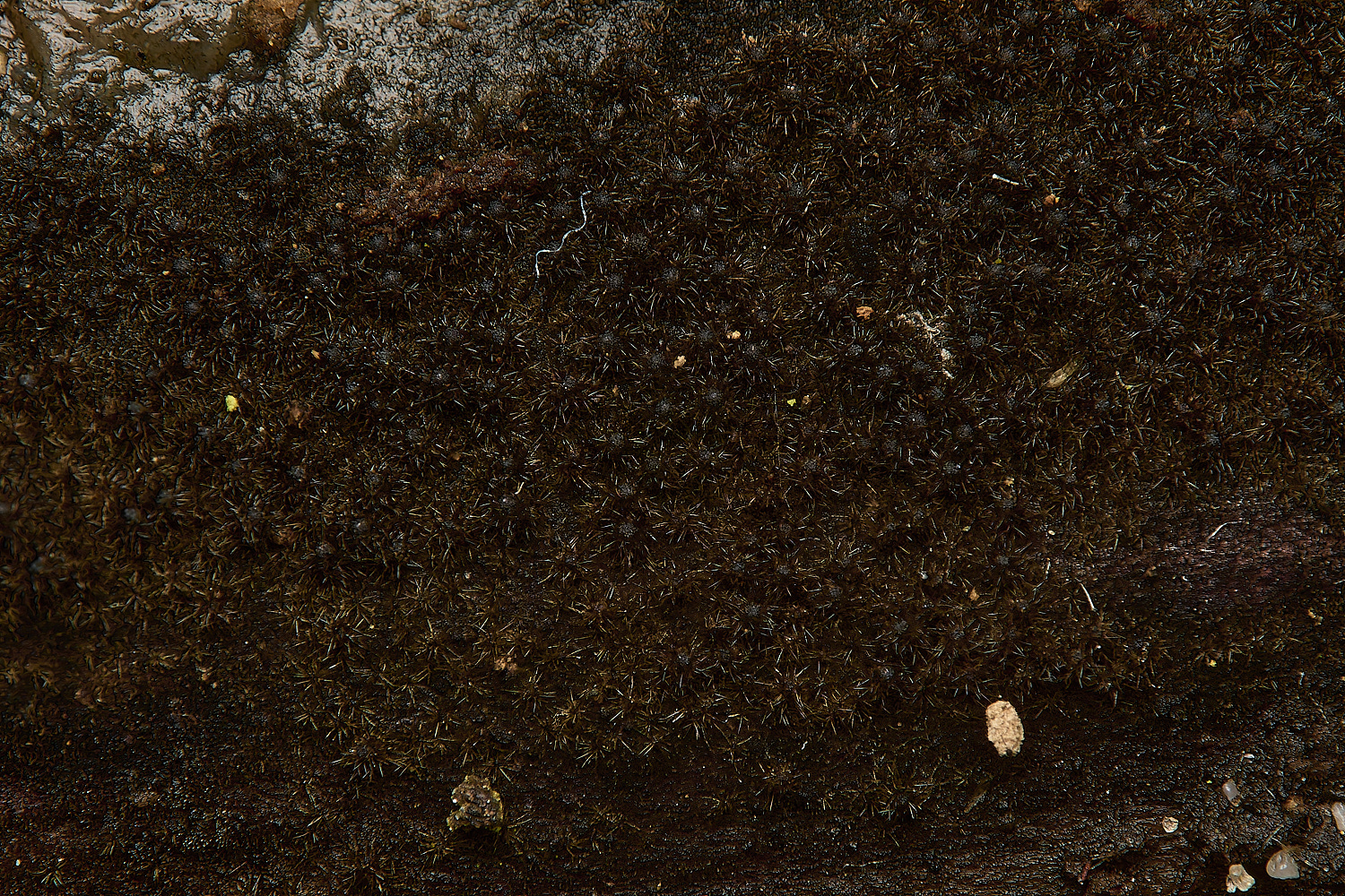
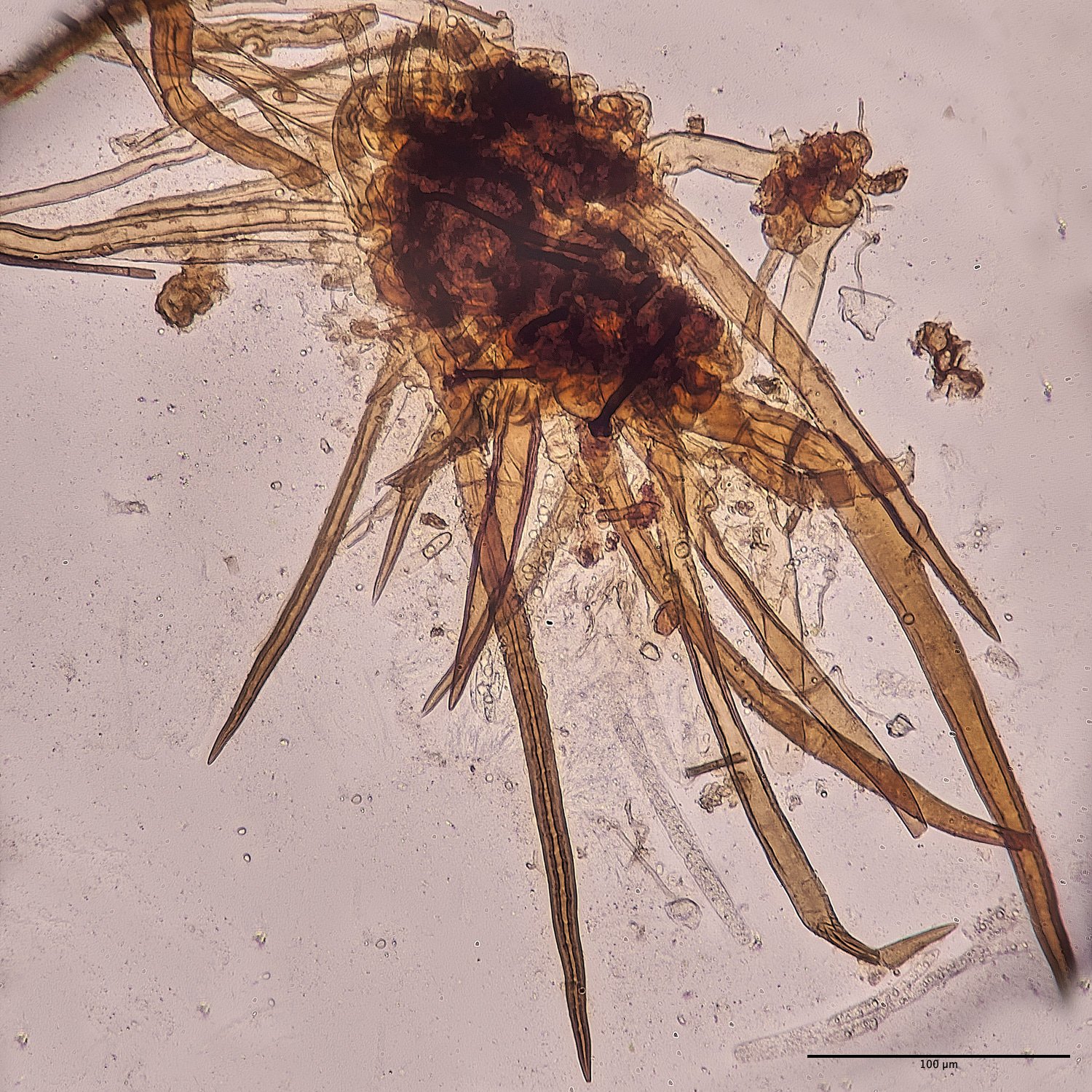
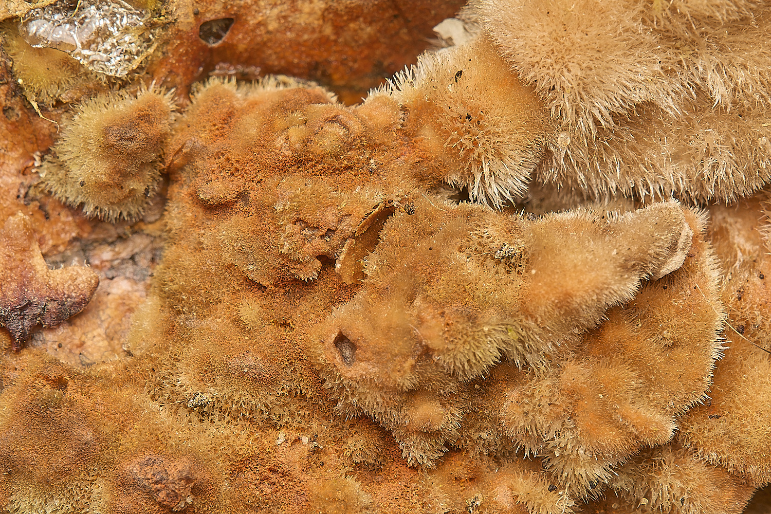
Small hairy fungus
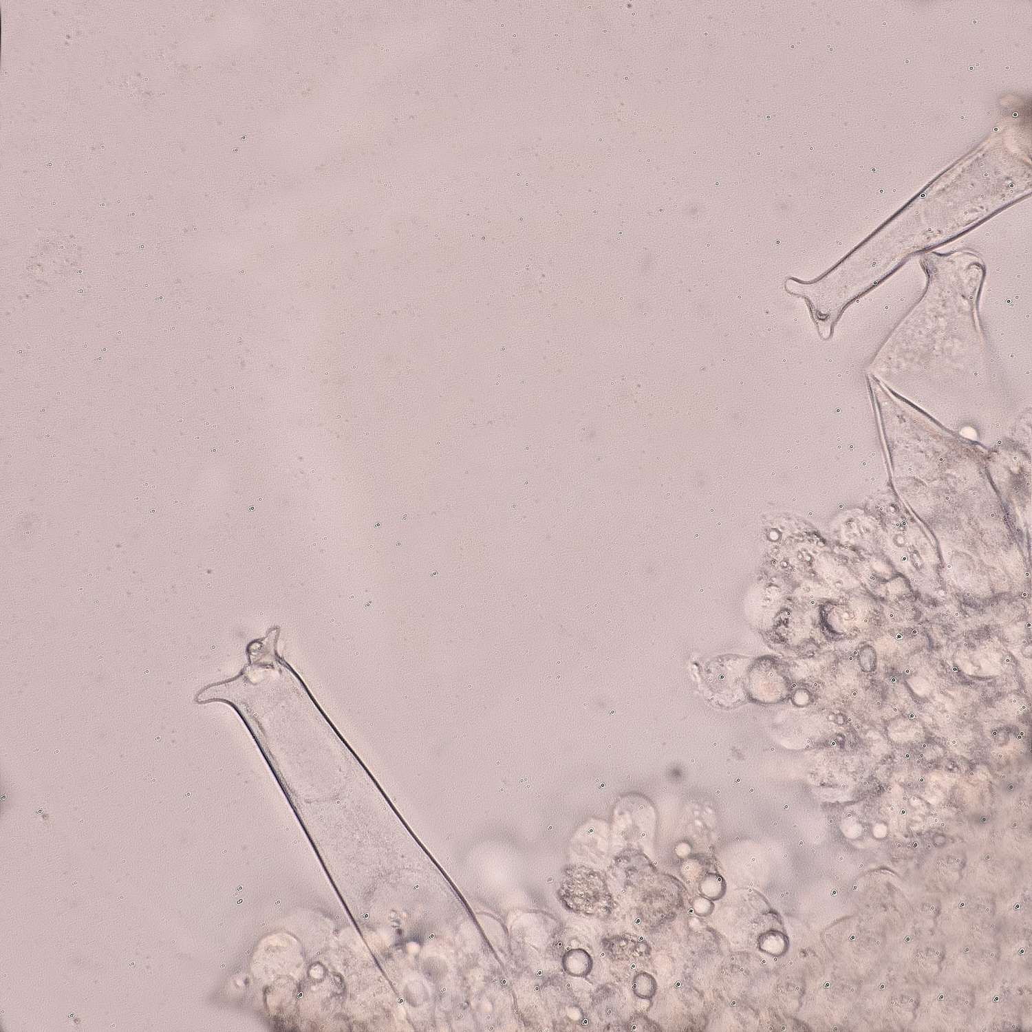
Lasiospherus hirsuta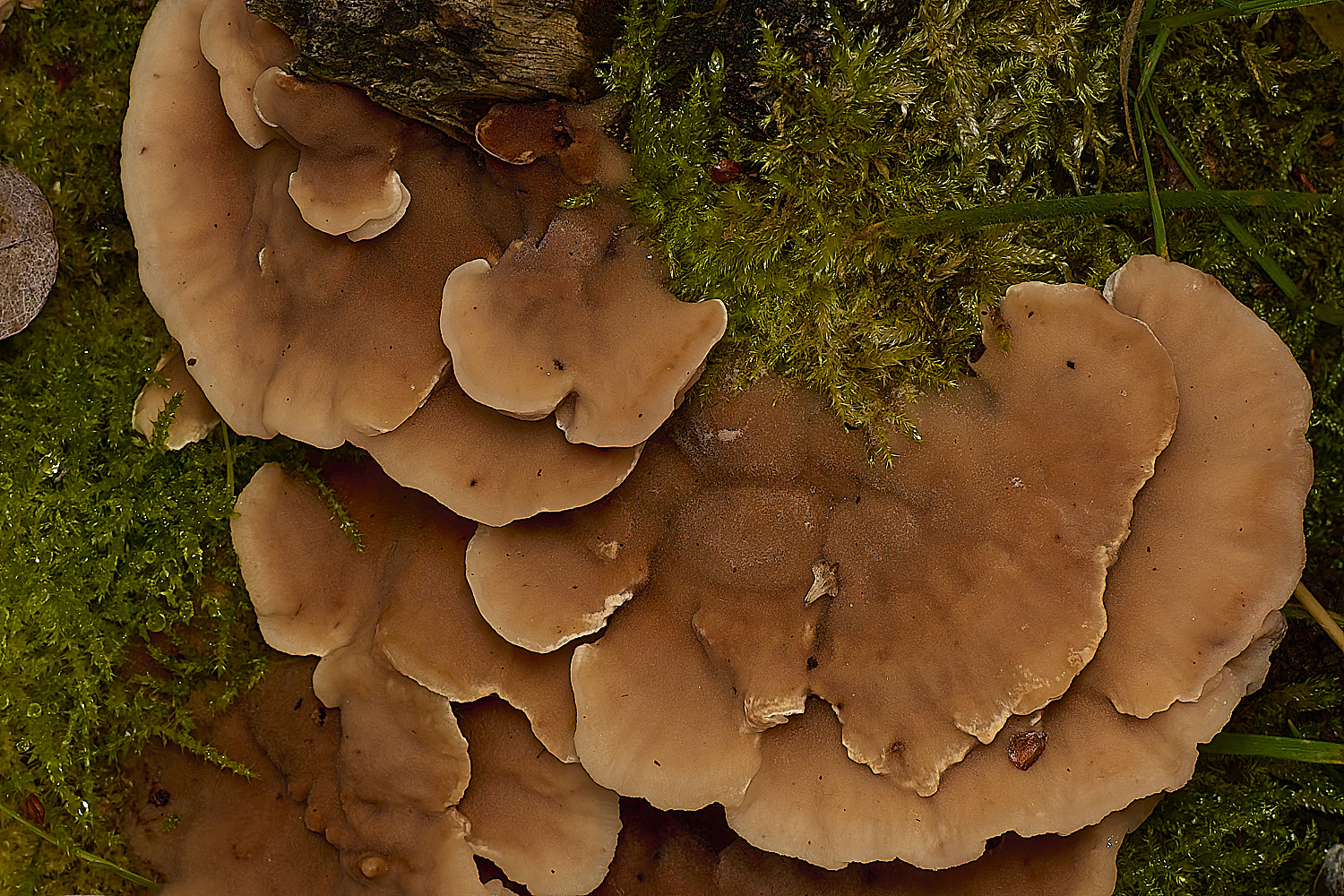
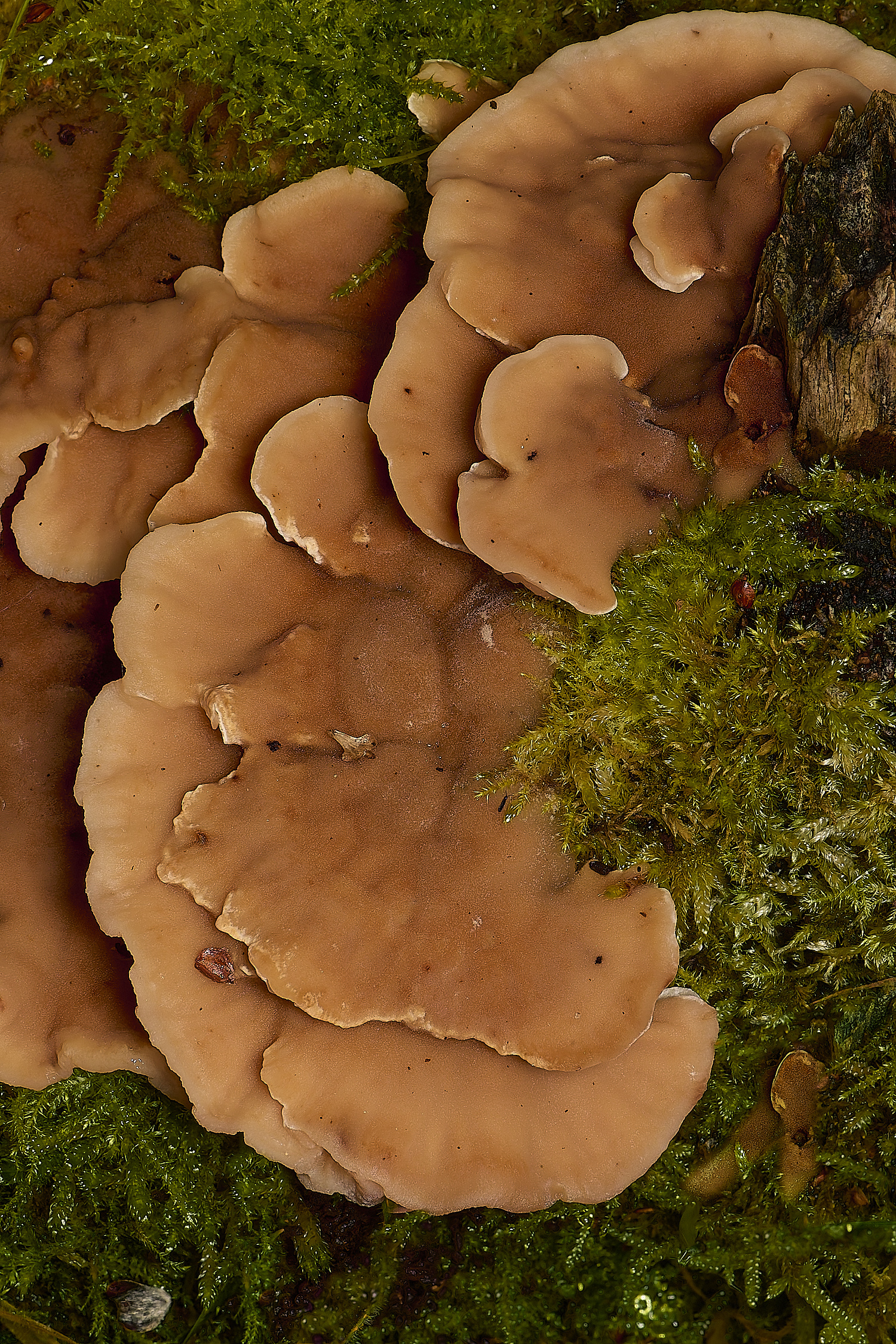
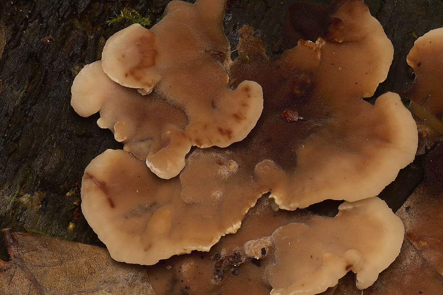
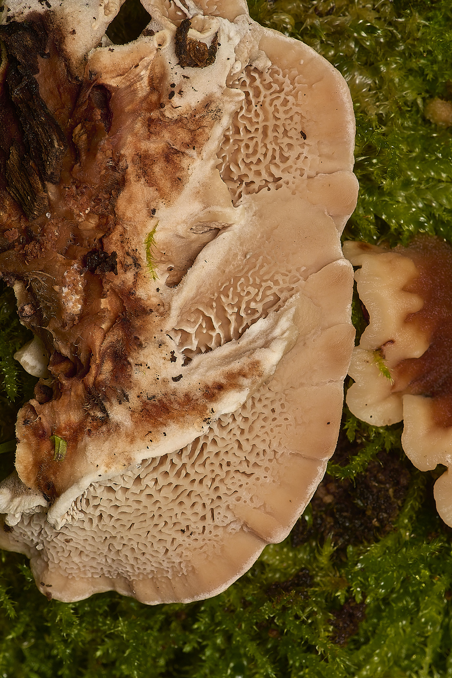
Blushing Rosette (Abortiporus biennis)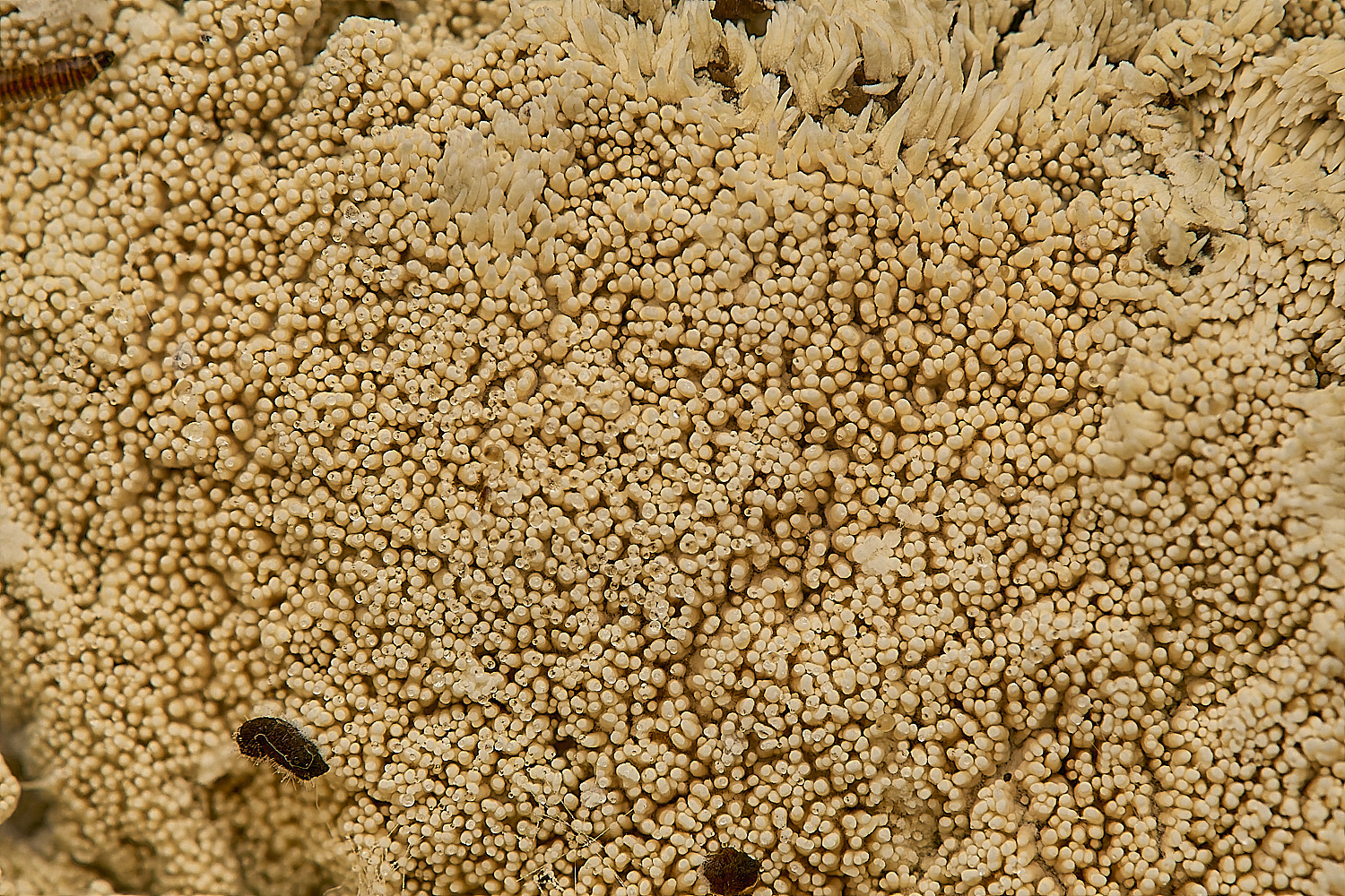
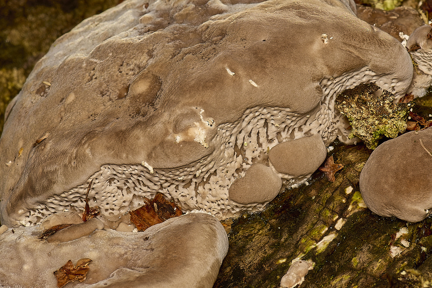
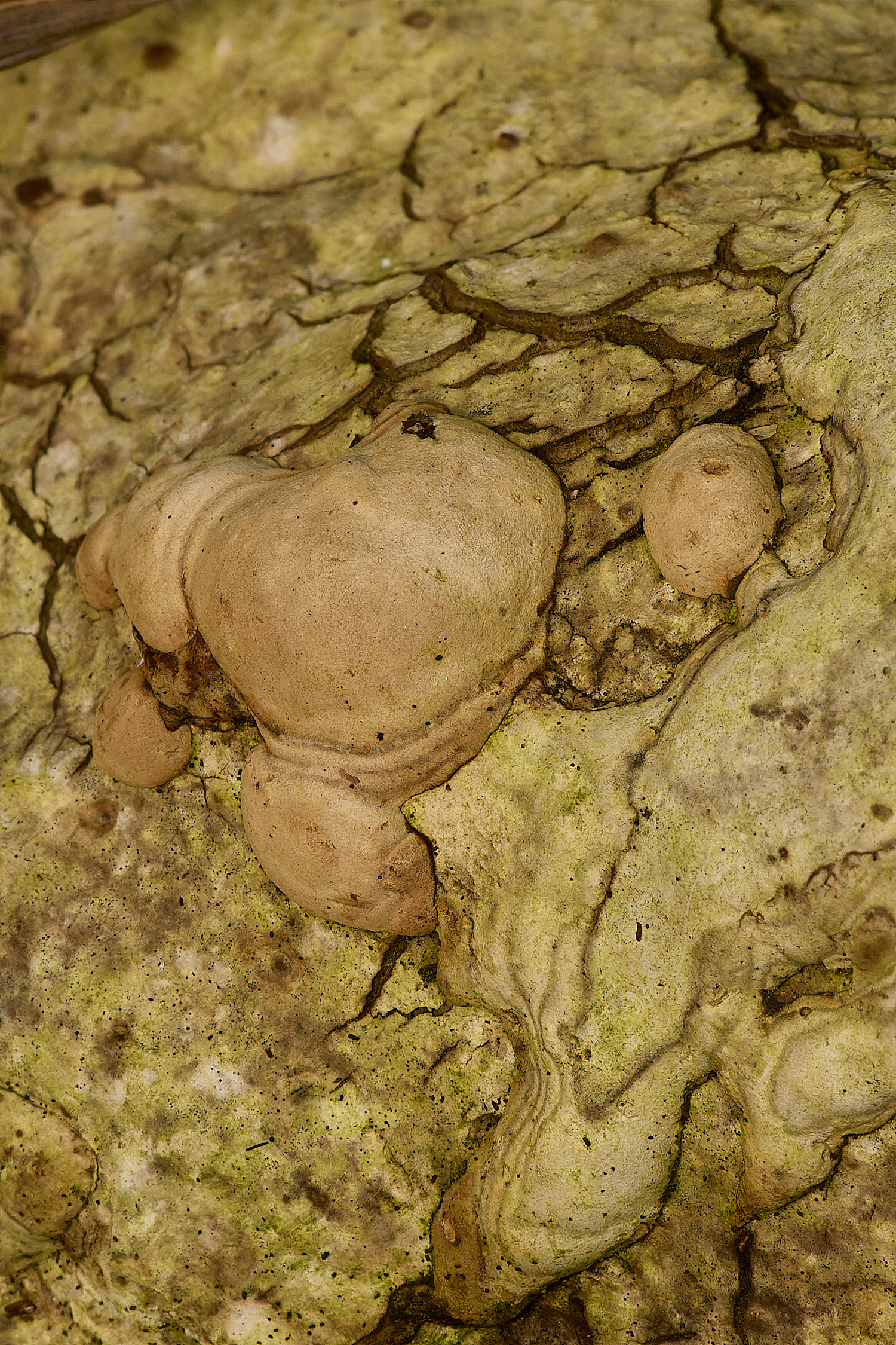
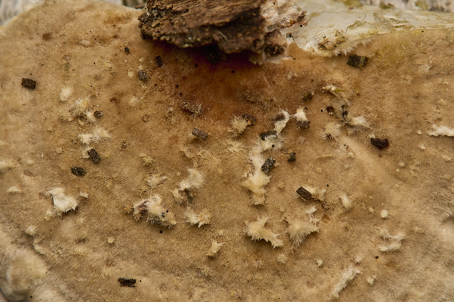
Toothed Fungus Sp (Trechispora farinacea) growing on a Hoof Fungus (Fomes fometarius)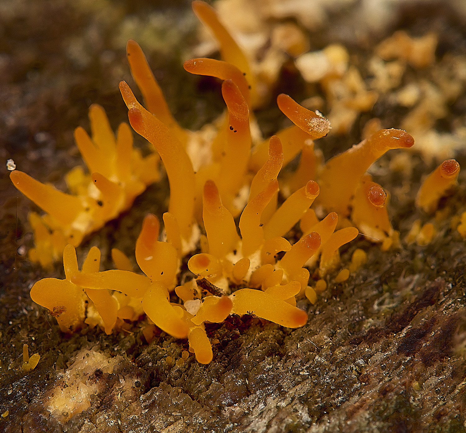
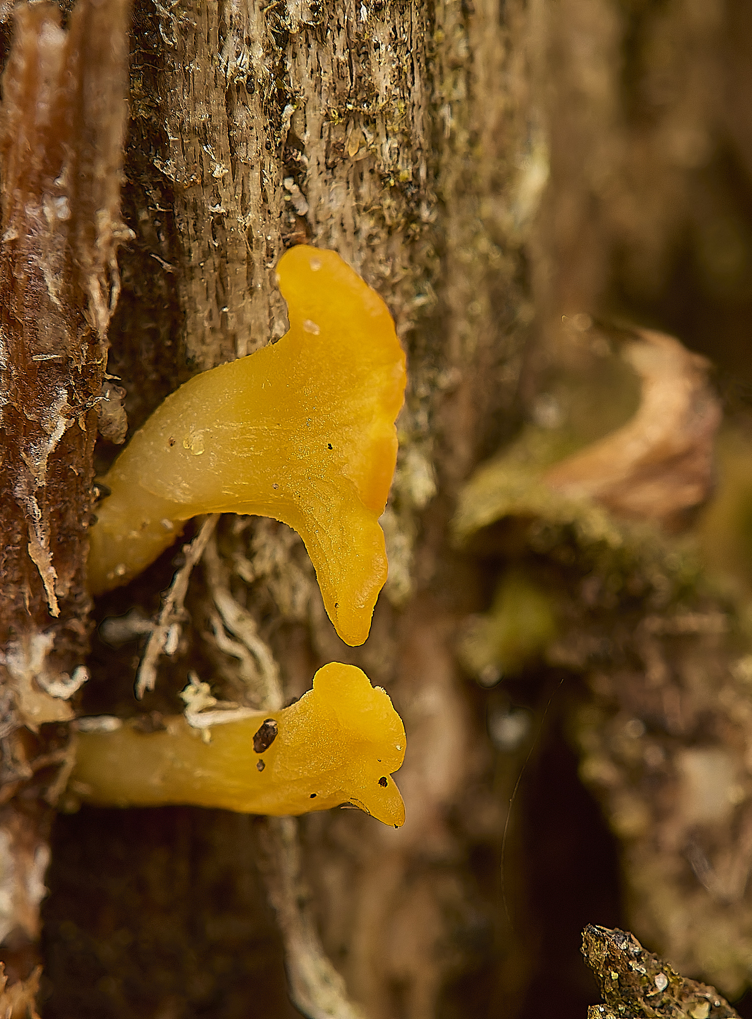
Small Stagshorn (Calocera cornea)
Wrinkled Crust (Phlebia radiata)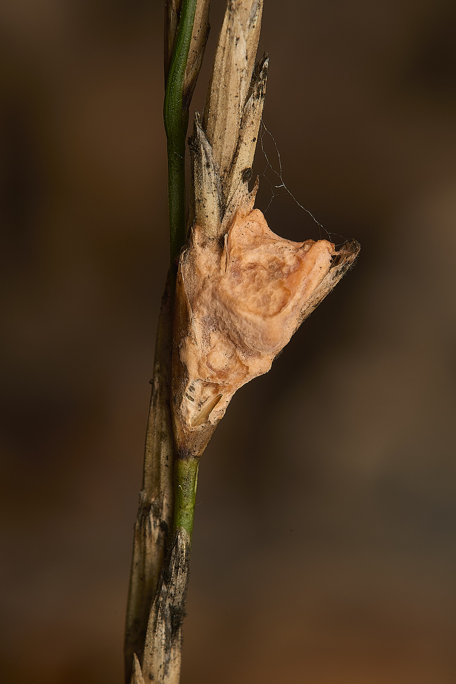
The fusarium state of Giberella zeae (Fusarium gramineum)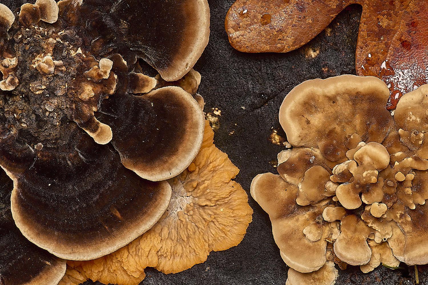
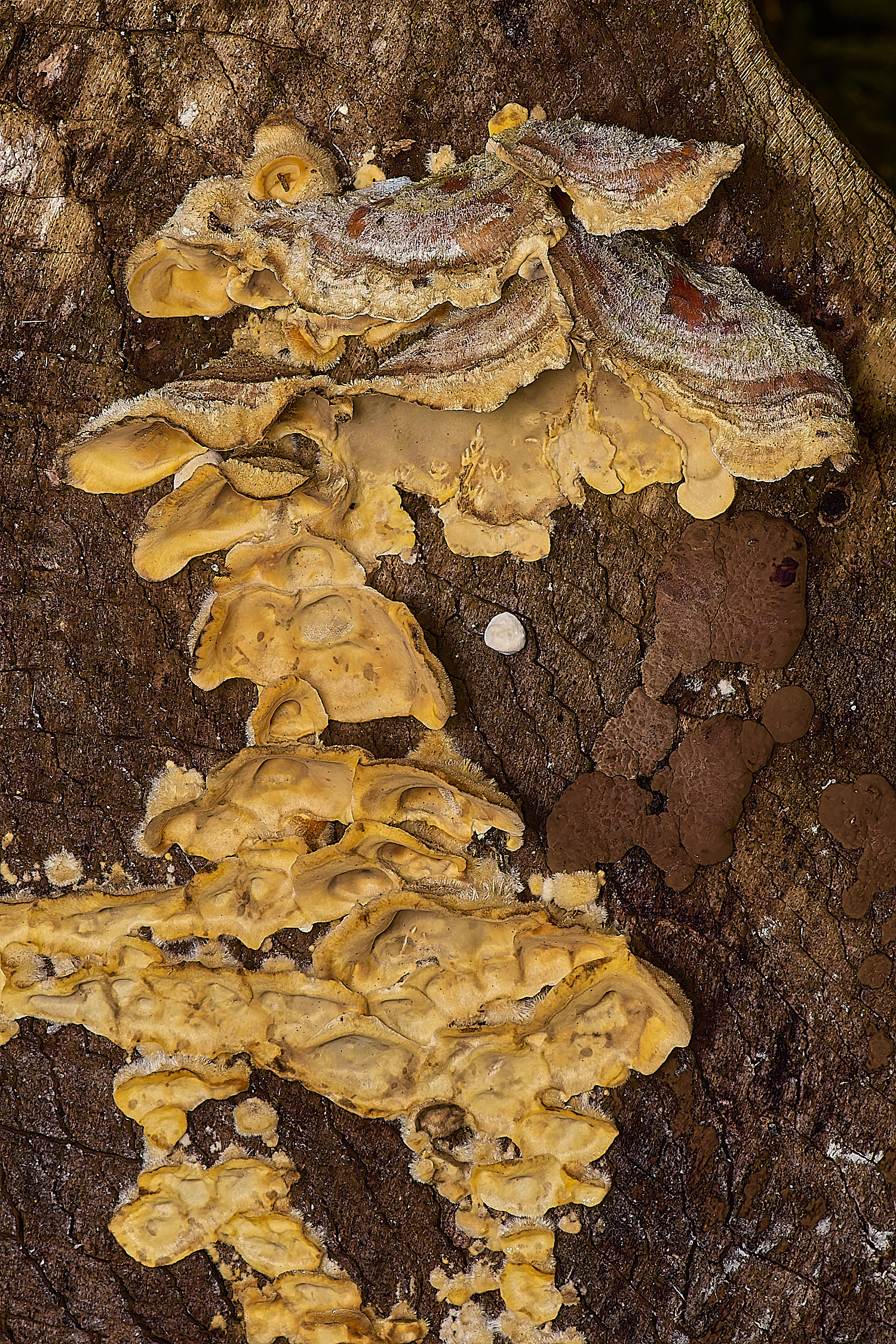
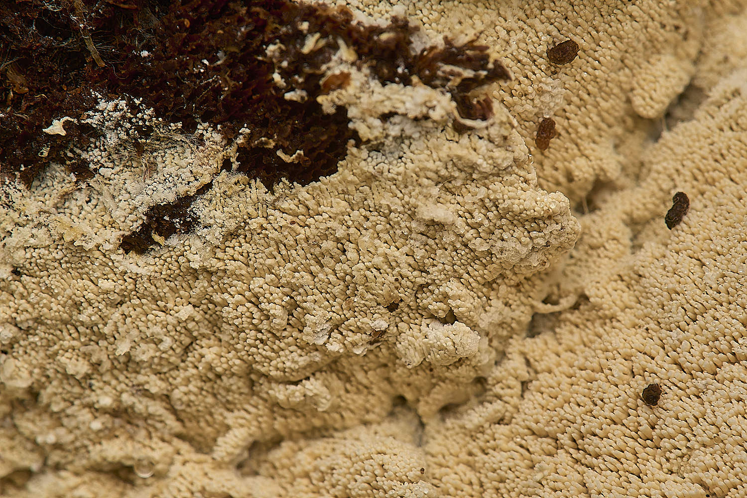
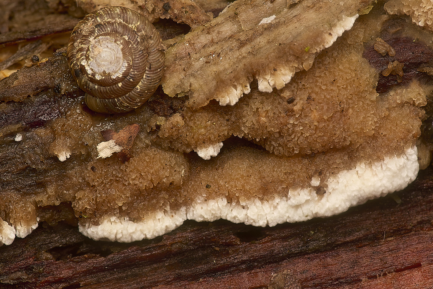
Mixed TurKey Tail, Wrinkled Crust & Blushing Rosette
Fly Agaric (Amanita muscaria)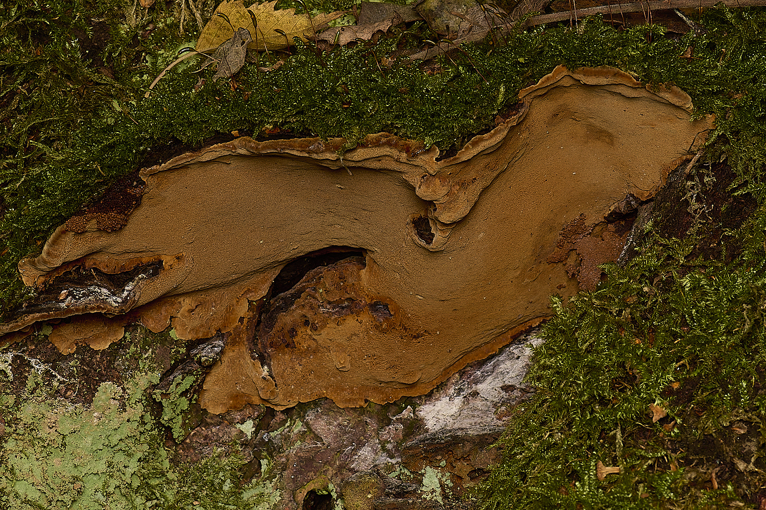
?
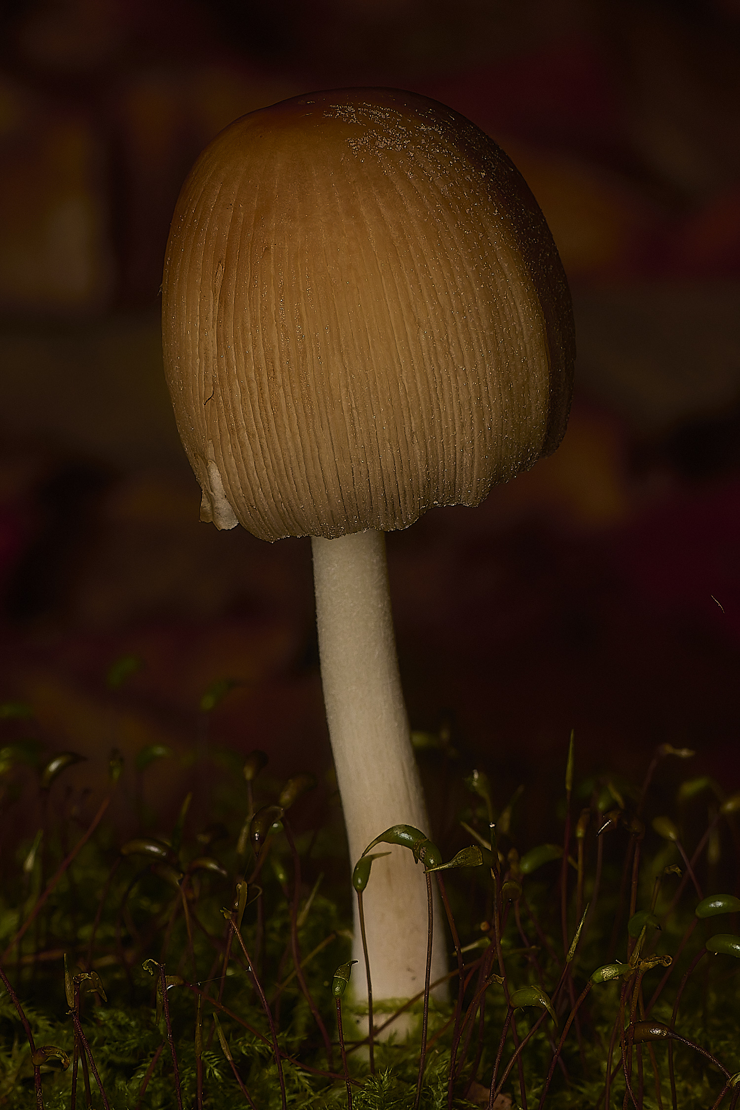
Glistening Inkcap (Coprinellus micaceus)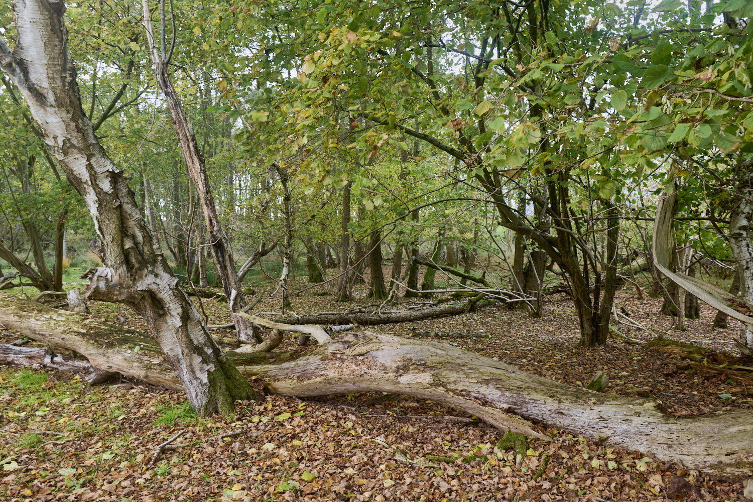
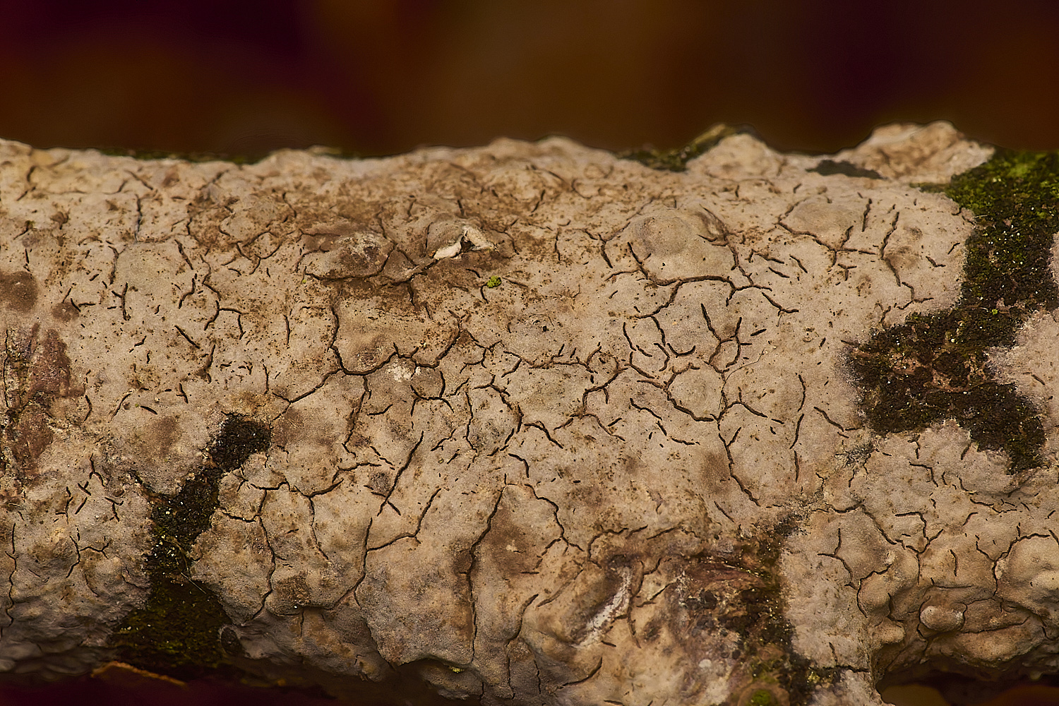
Waxy Crust on Hawthorn (Crataegus monogyna)
Vuilleminia cystidata 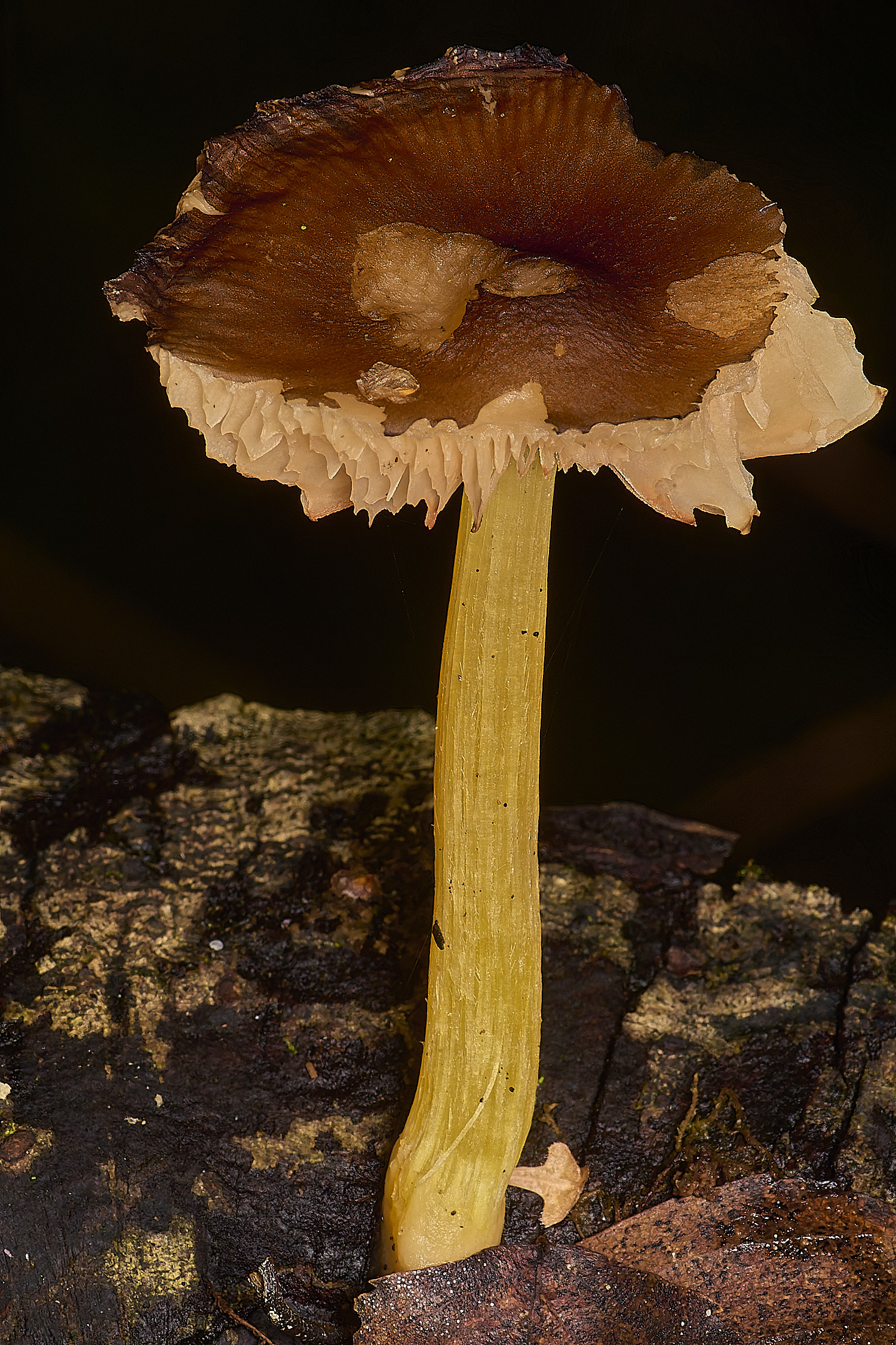
Goldleaf Shield (Pluteus romellii)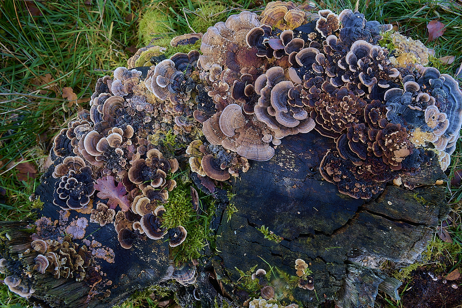
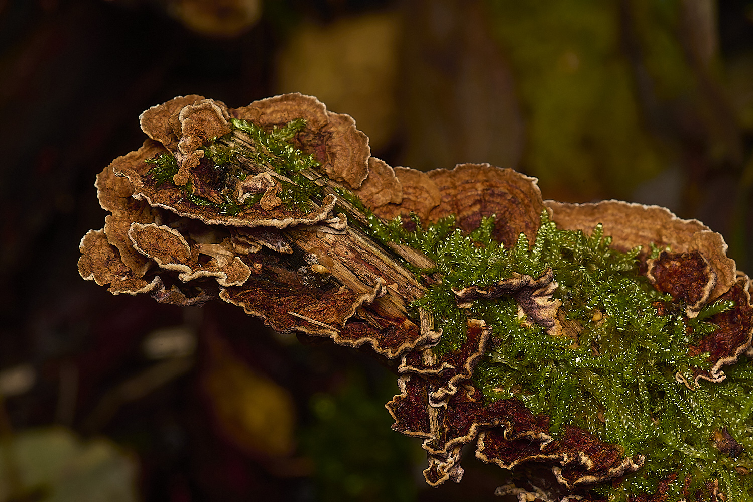
A mixed stump of Turkey Tail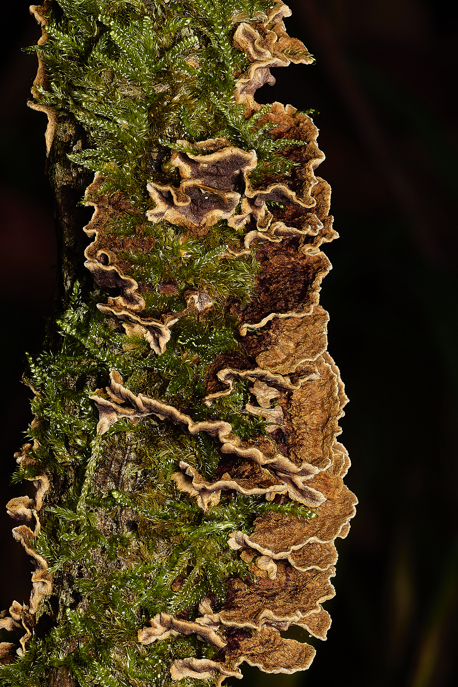
Hymenochaete tabacina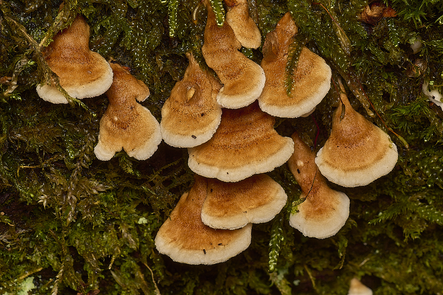
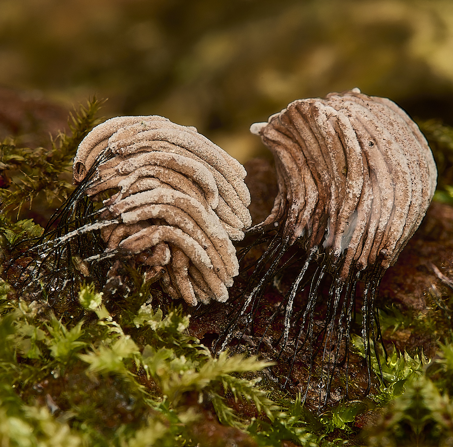
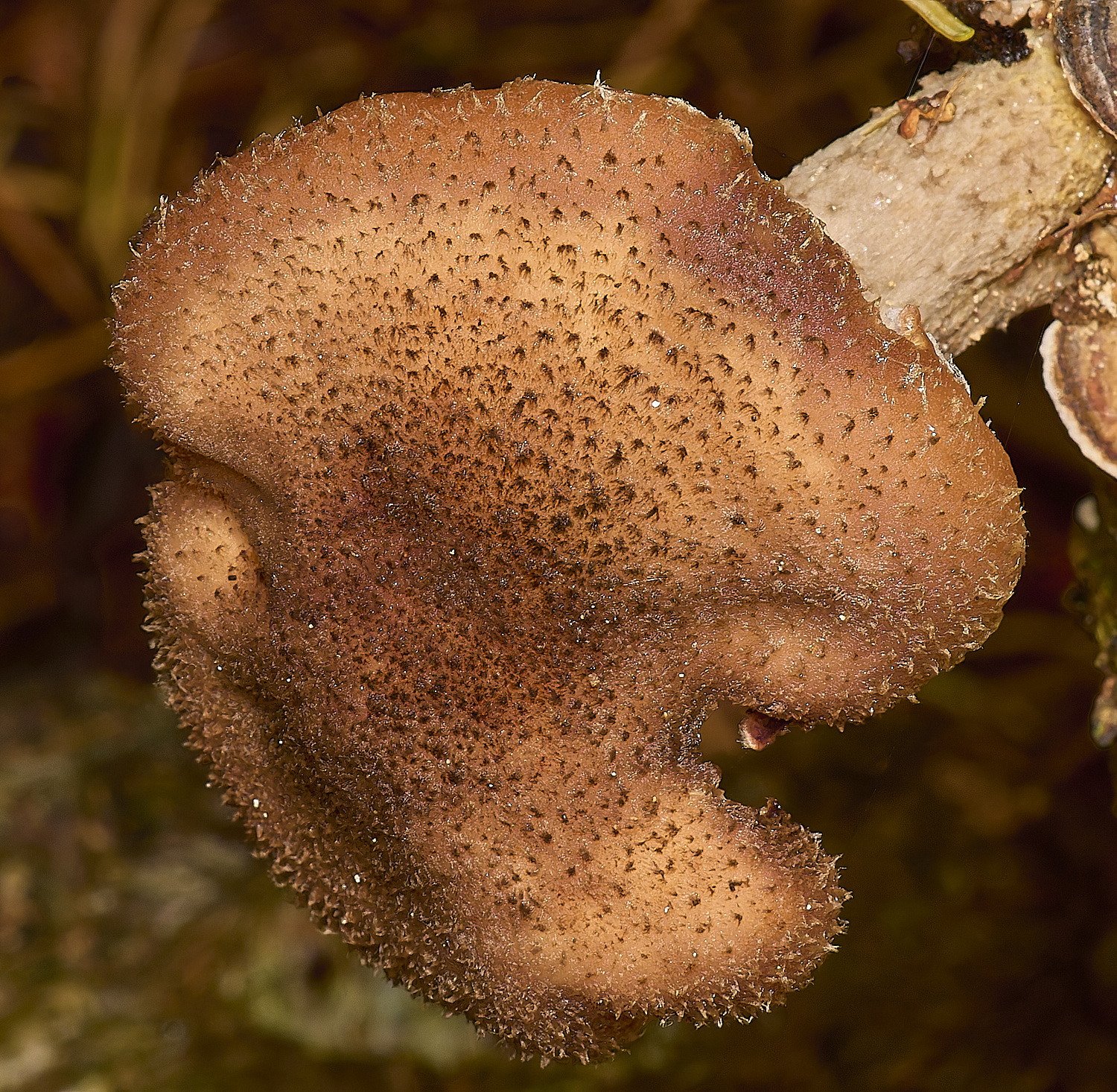
Crimpgill (Plicatura crispa)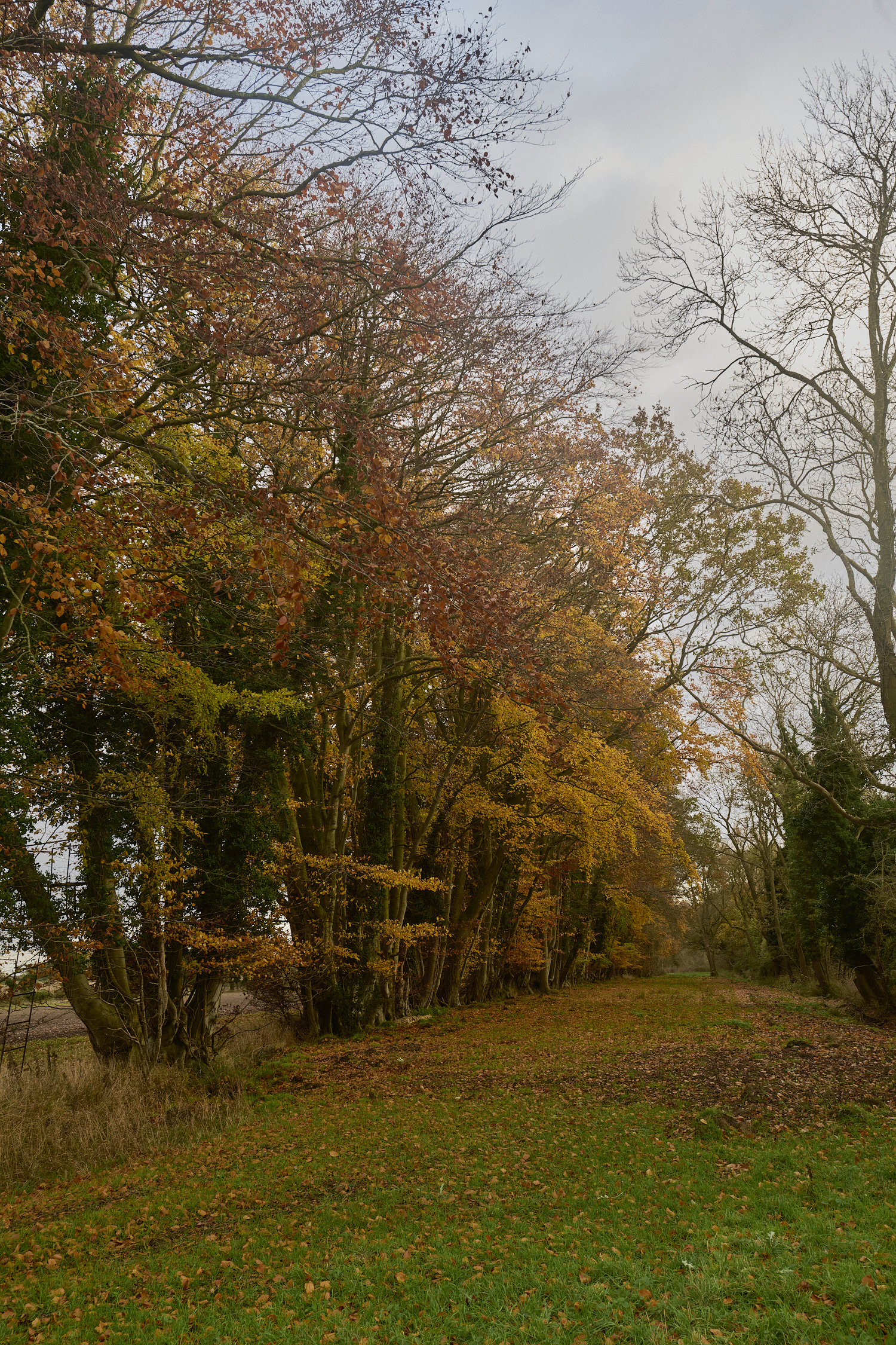
Beech (Fagus sylvatica)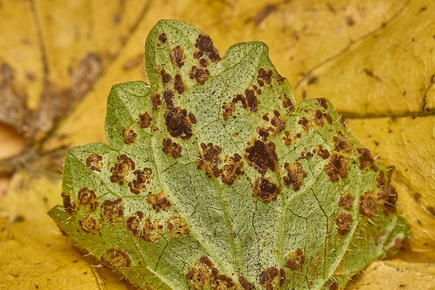
A rust (Puccinia glechomatis) on Ground Ivy (Glechoma hederacea)
Felbrigg
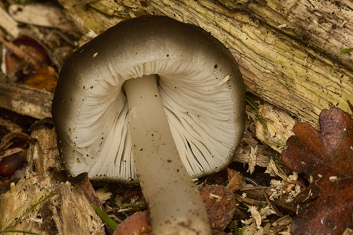
Burgundy Drop Bonnet (Mycena haematopus)?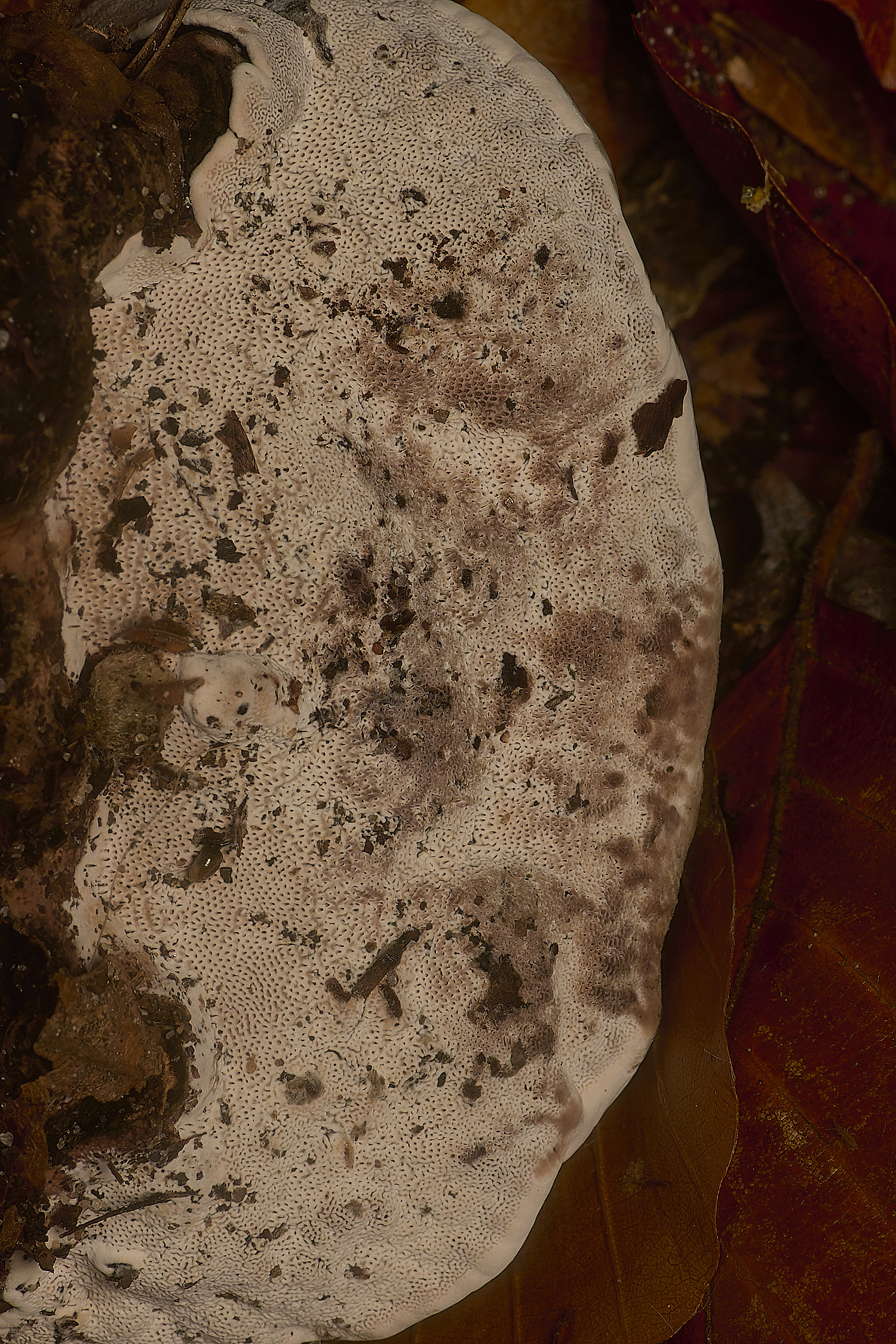
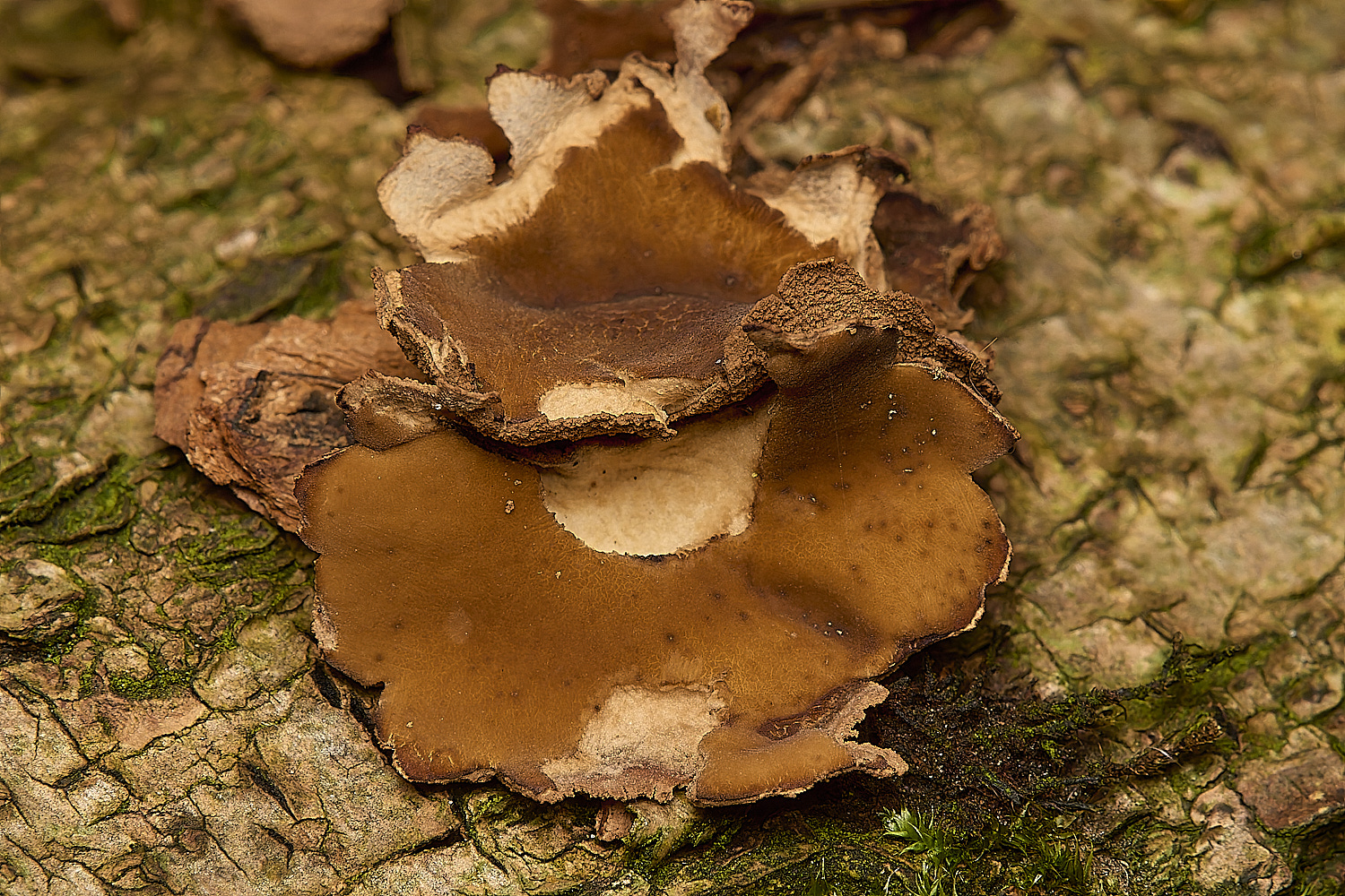
Blushing Bracket (Daedalopsis contragosa)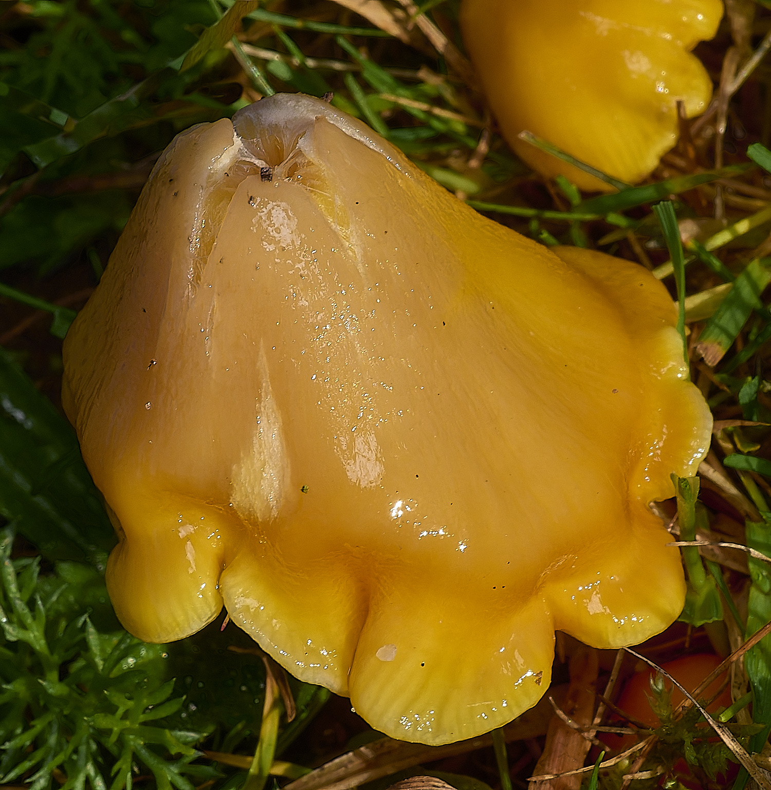
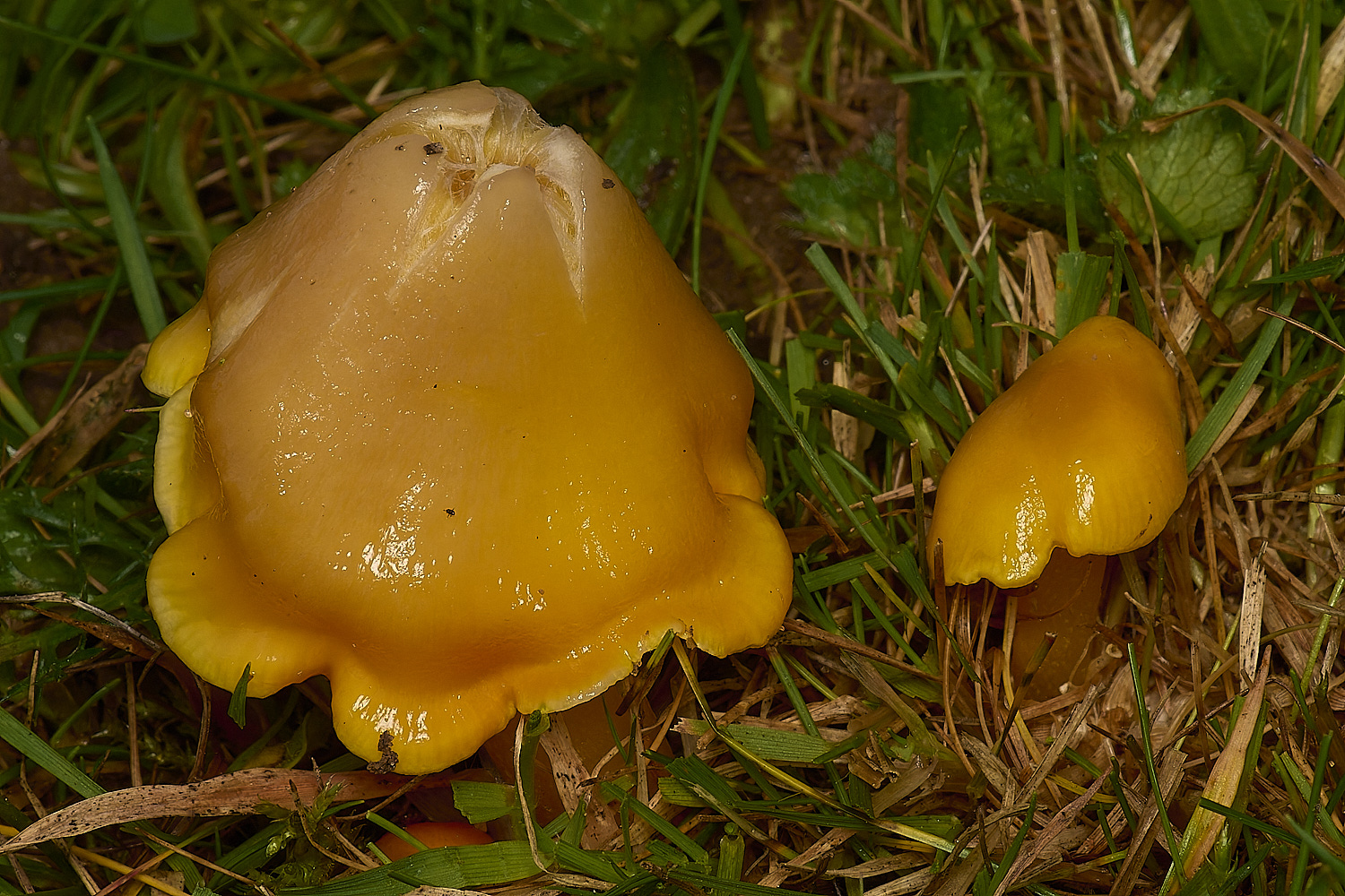
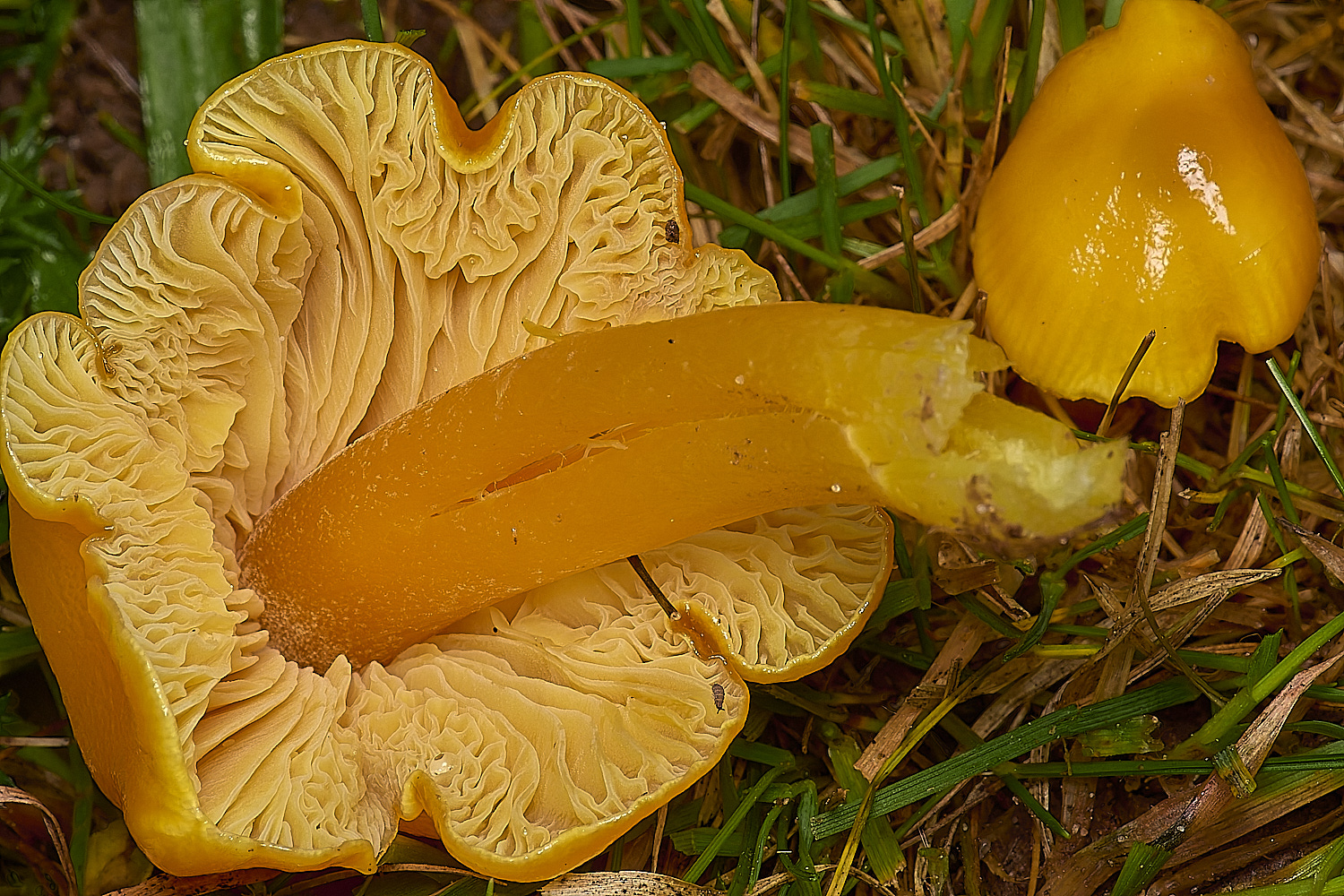
Golden Waxcap (Hydrocybe chlorophana)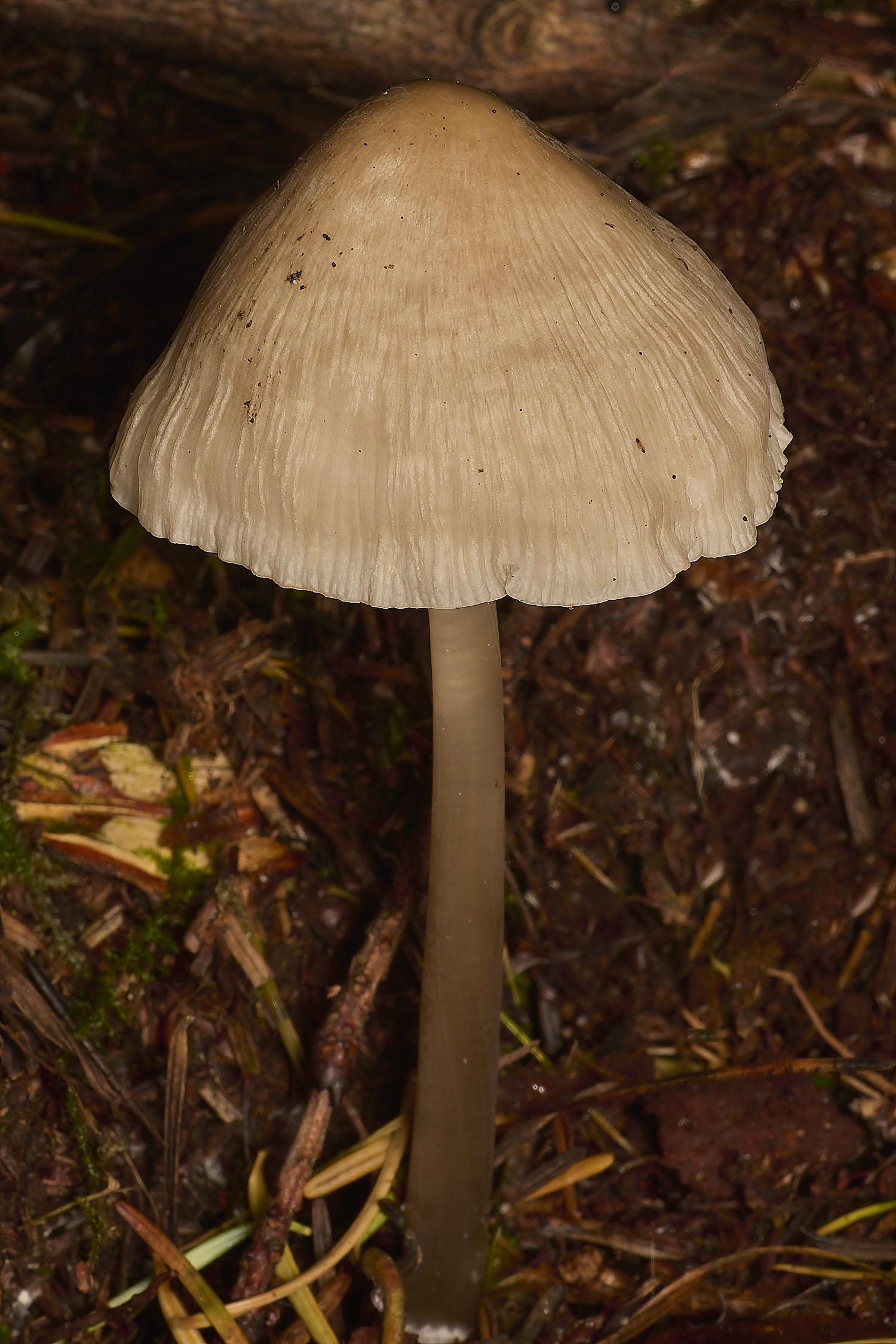
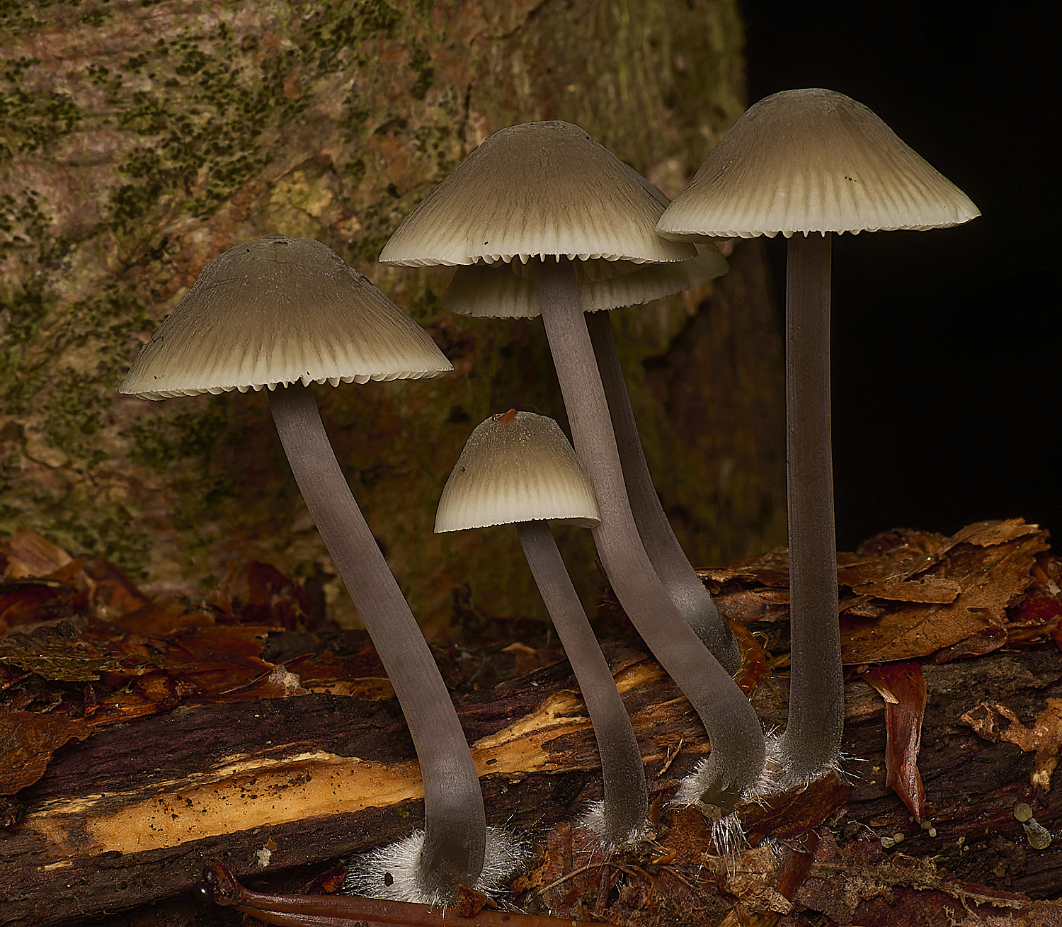
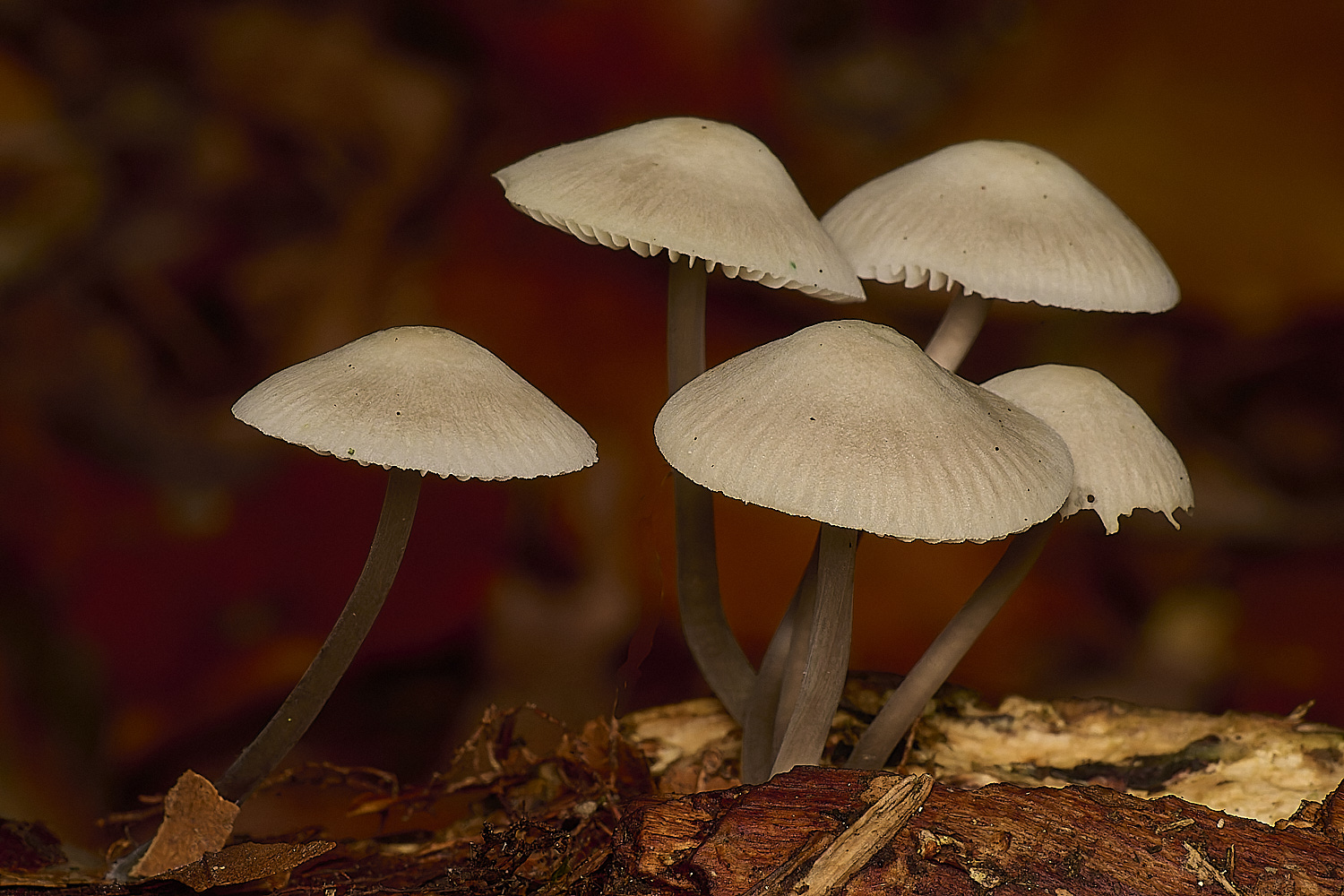
Common Bonnet (Mycena galericulata)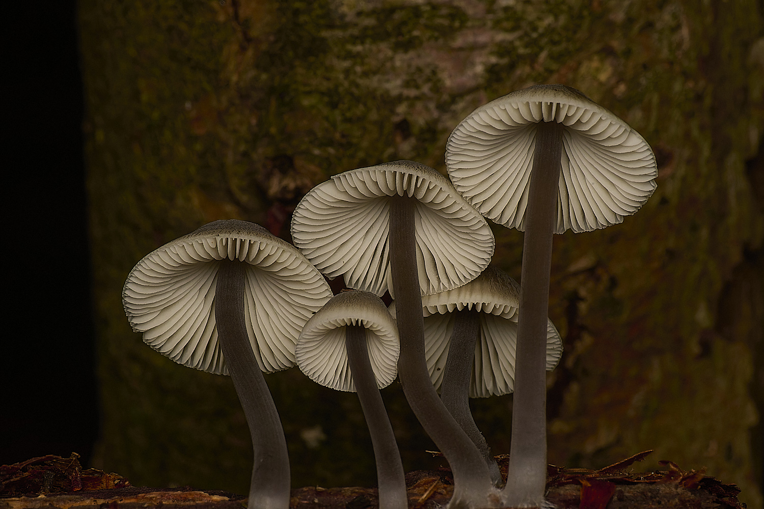
Mycena Sp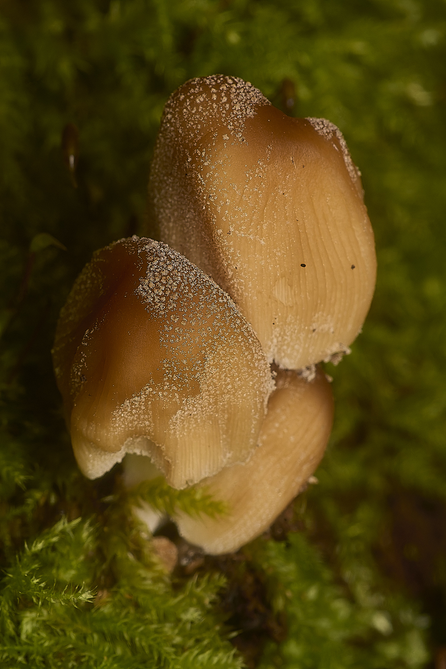
Glistening Inkcap (Coprinellus micaceus)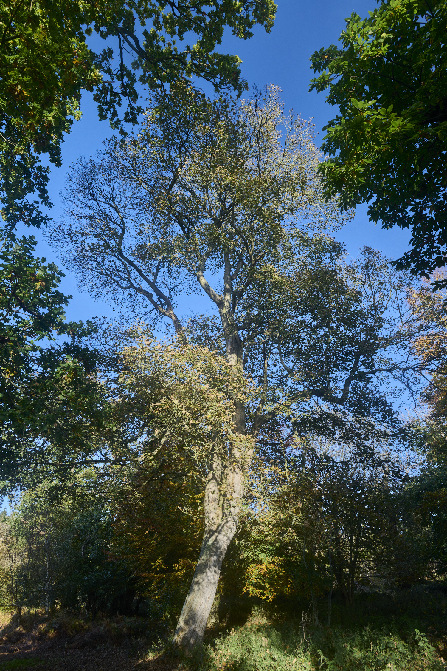
Sycamore (Acer psuedoplatanus)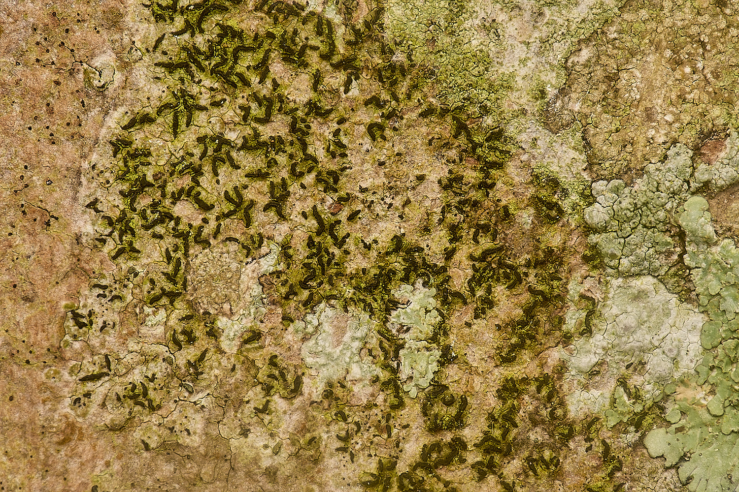
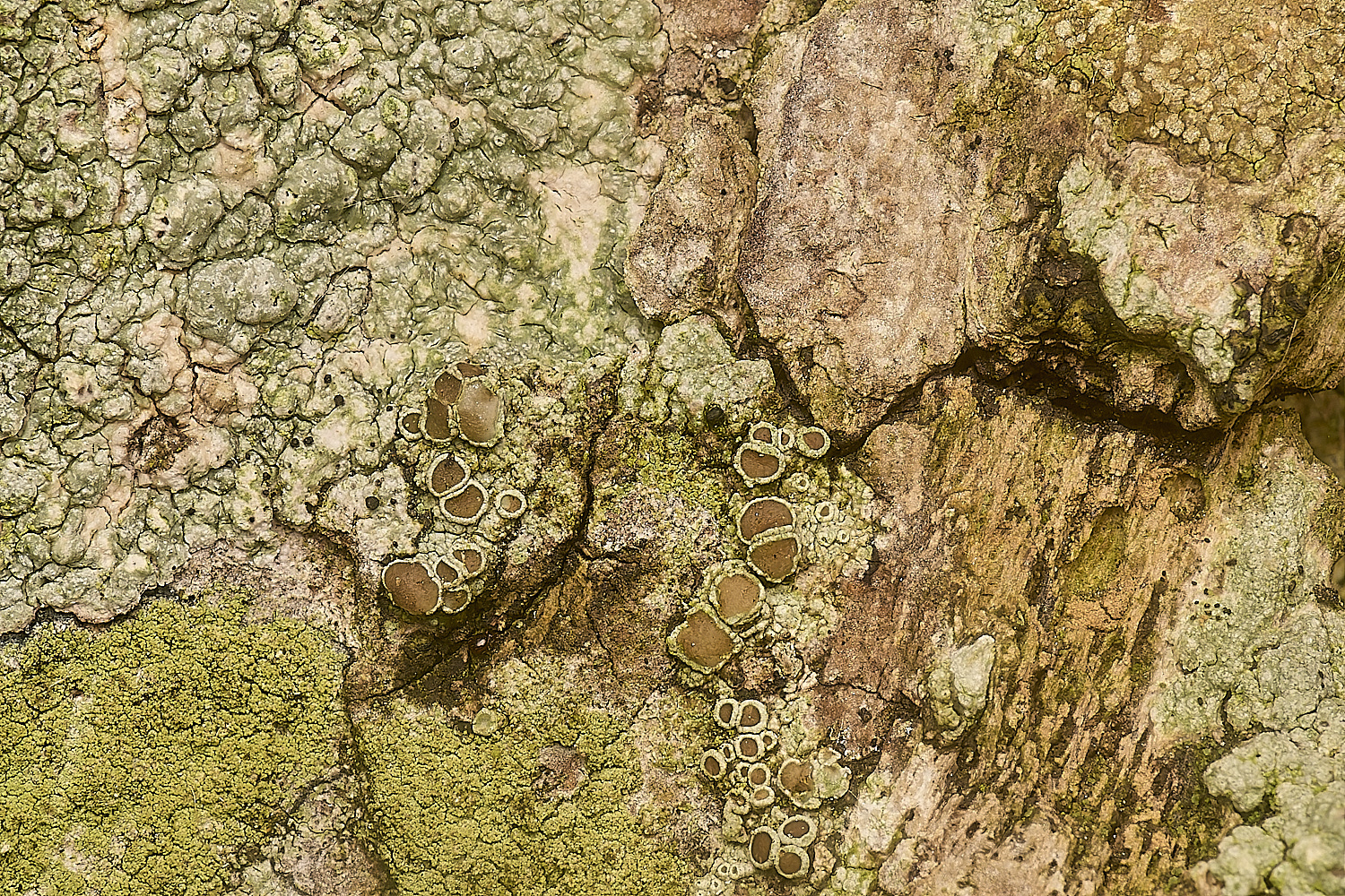
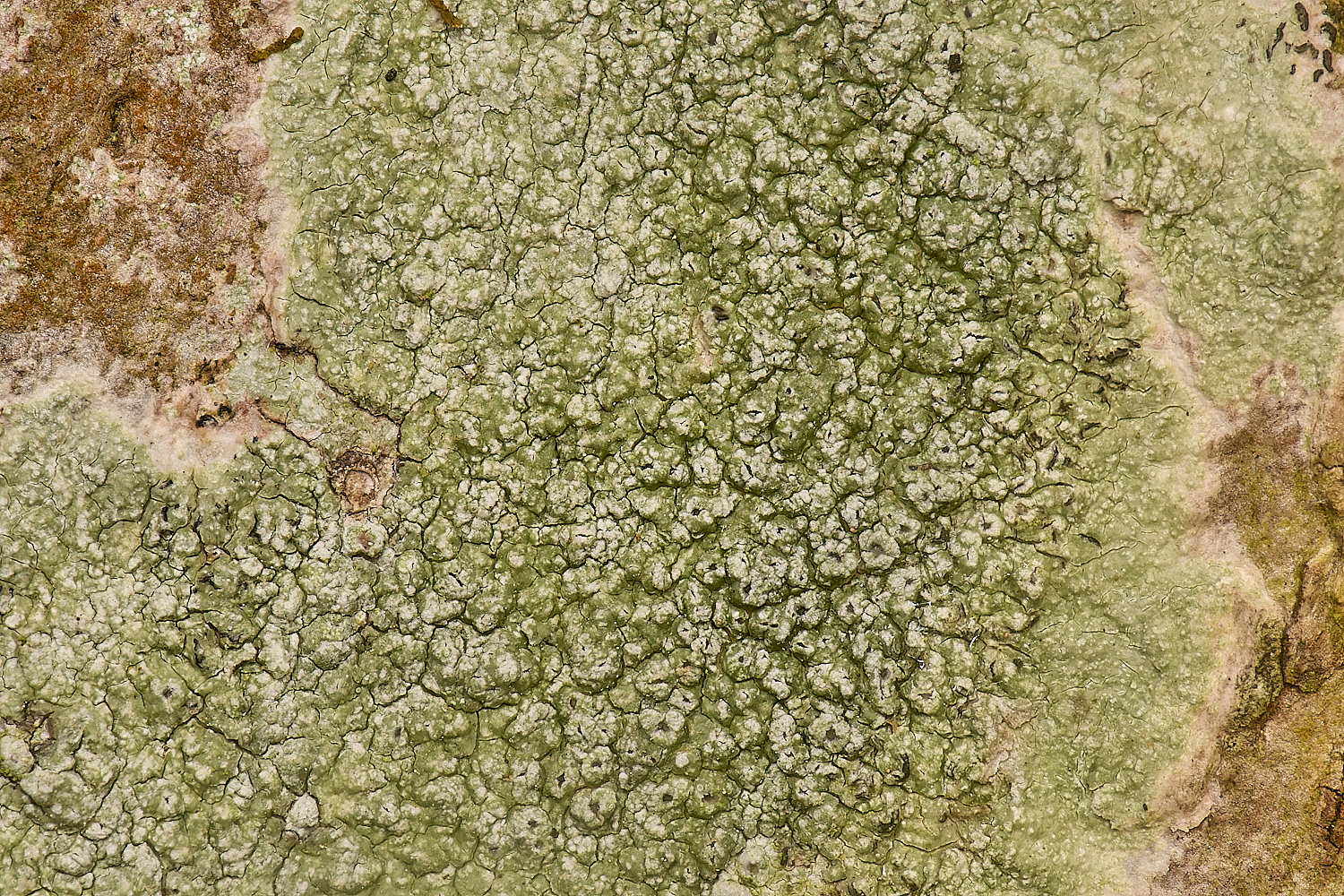
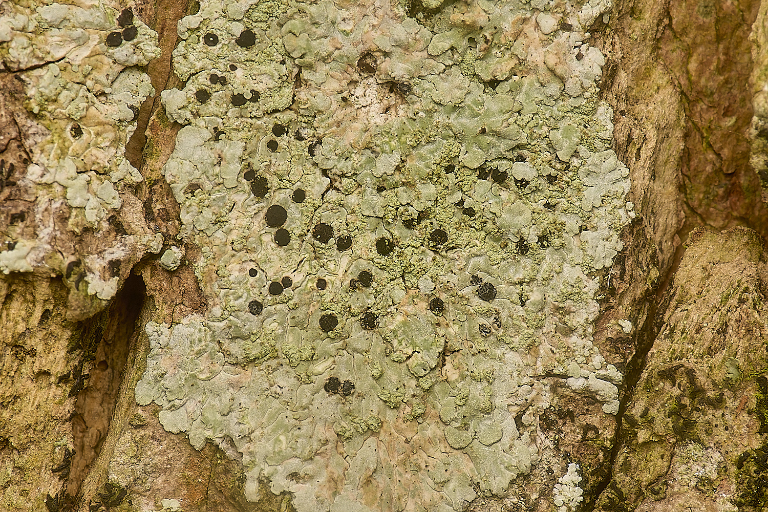
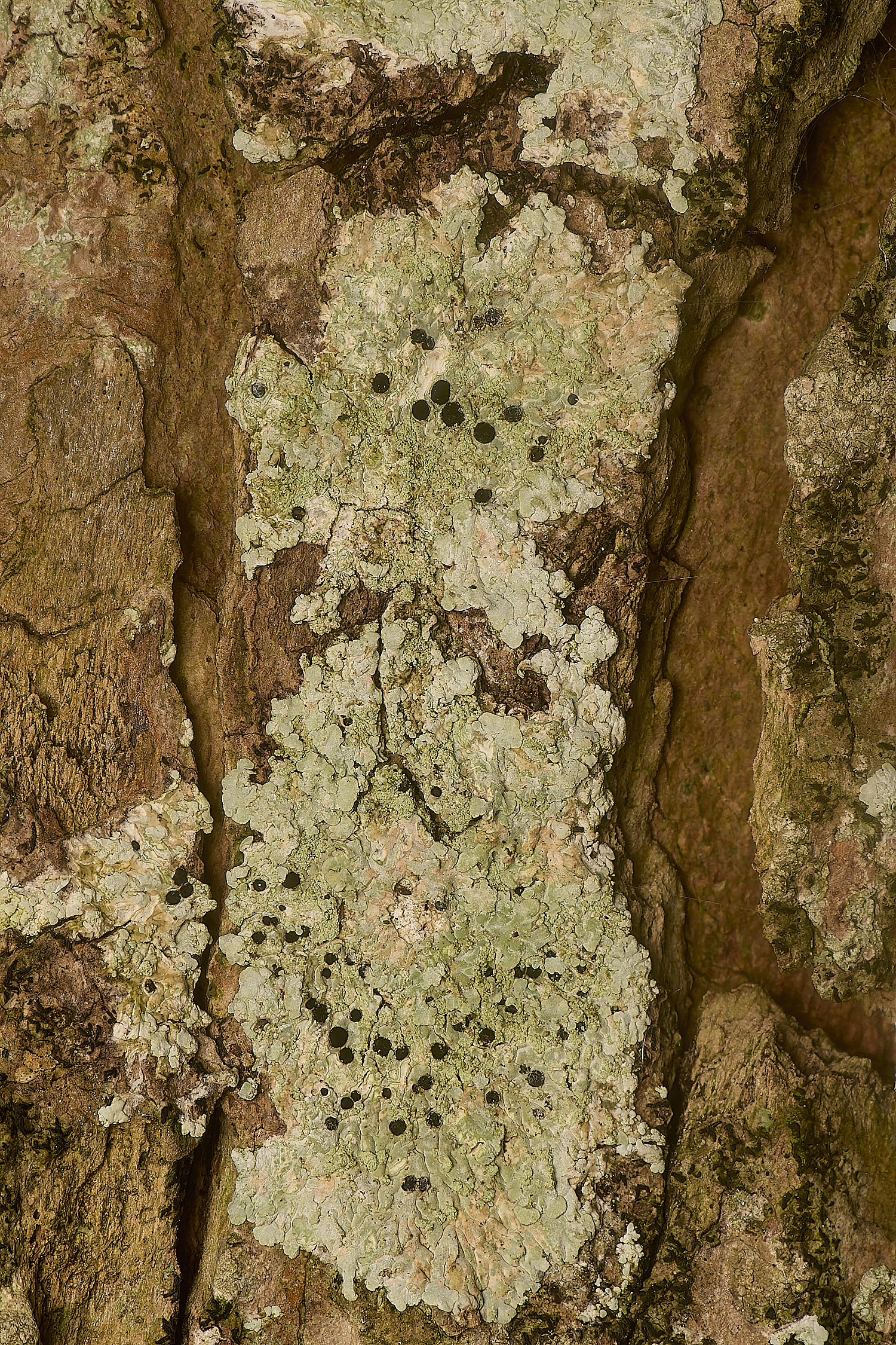
Lichen Sp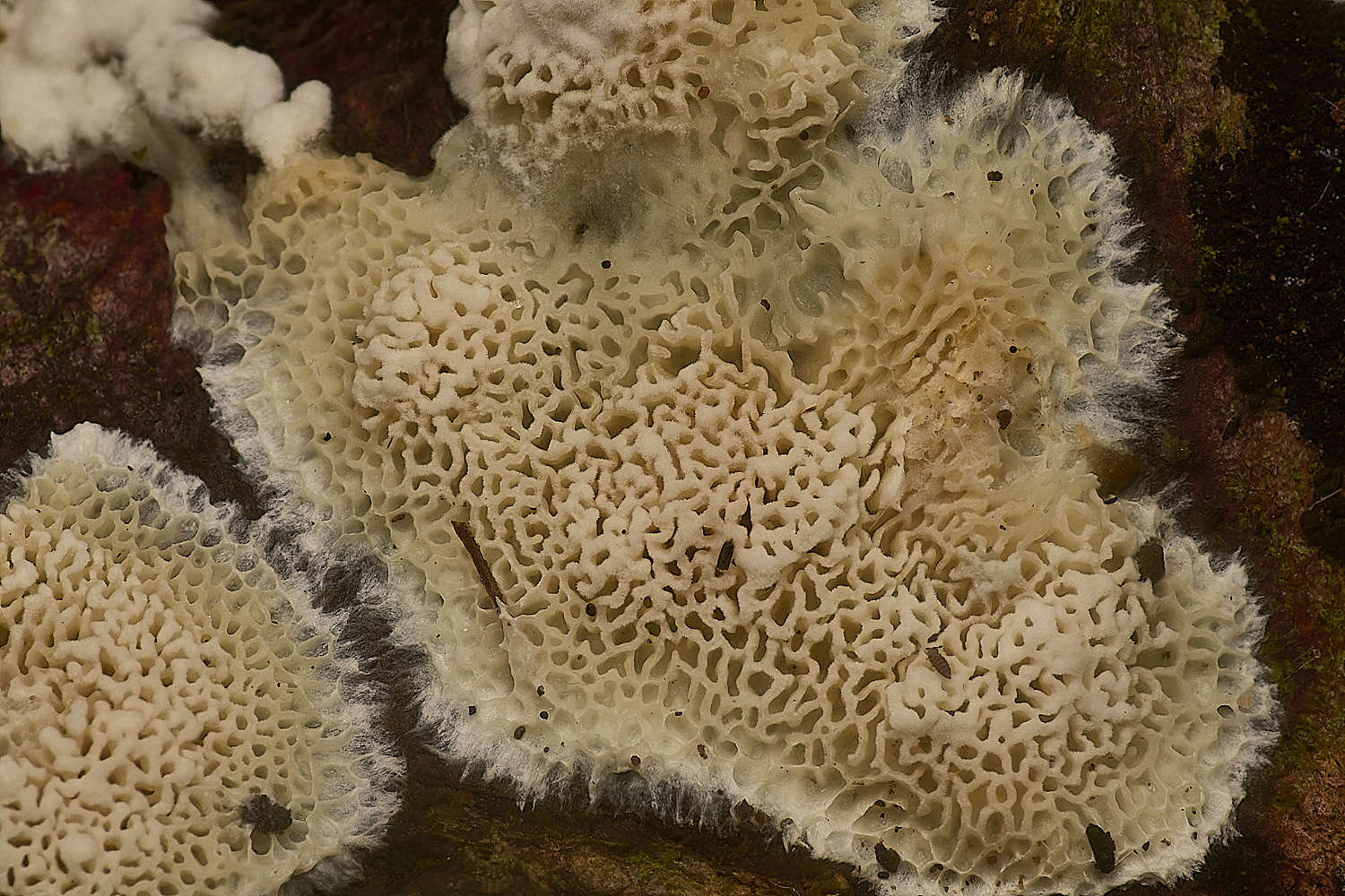
?
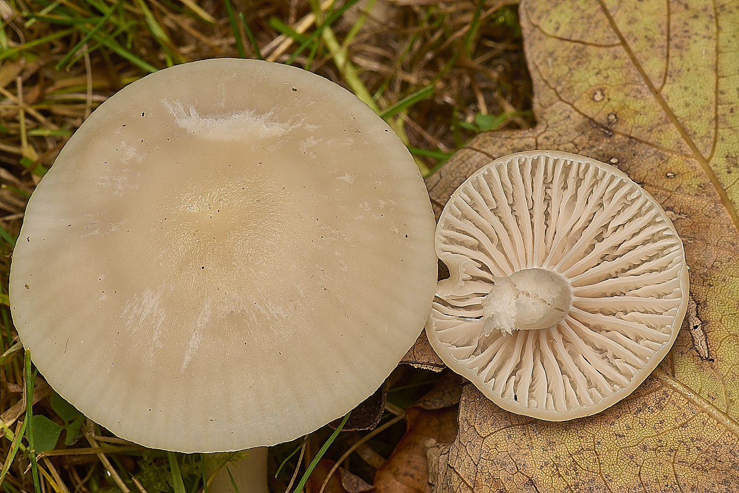
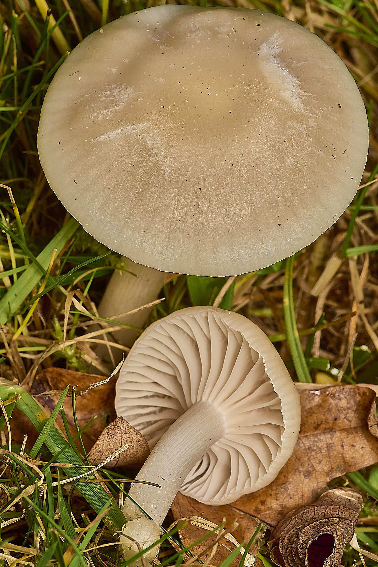
Meadow Waxcap (Cupophyllus pratensis)?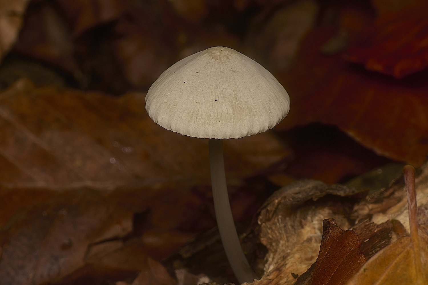

Sp?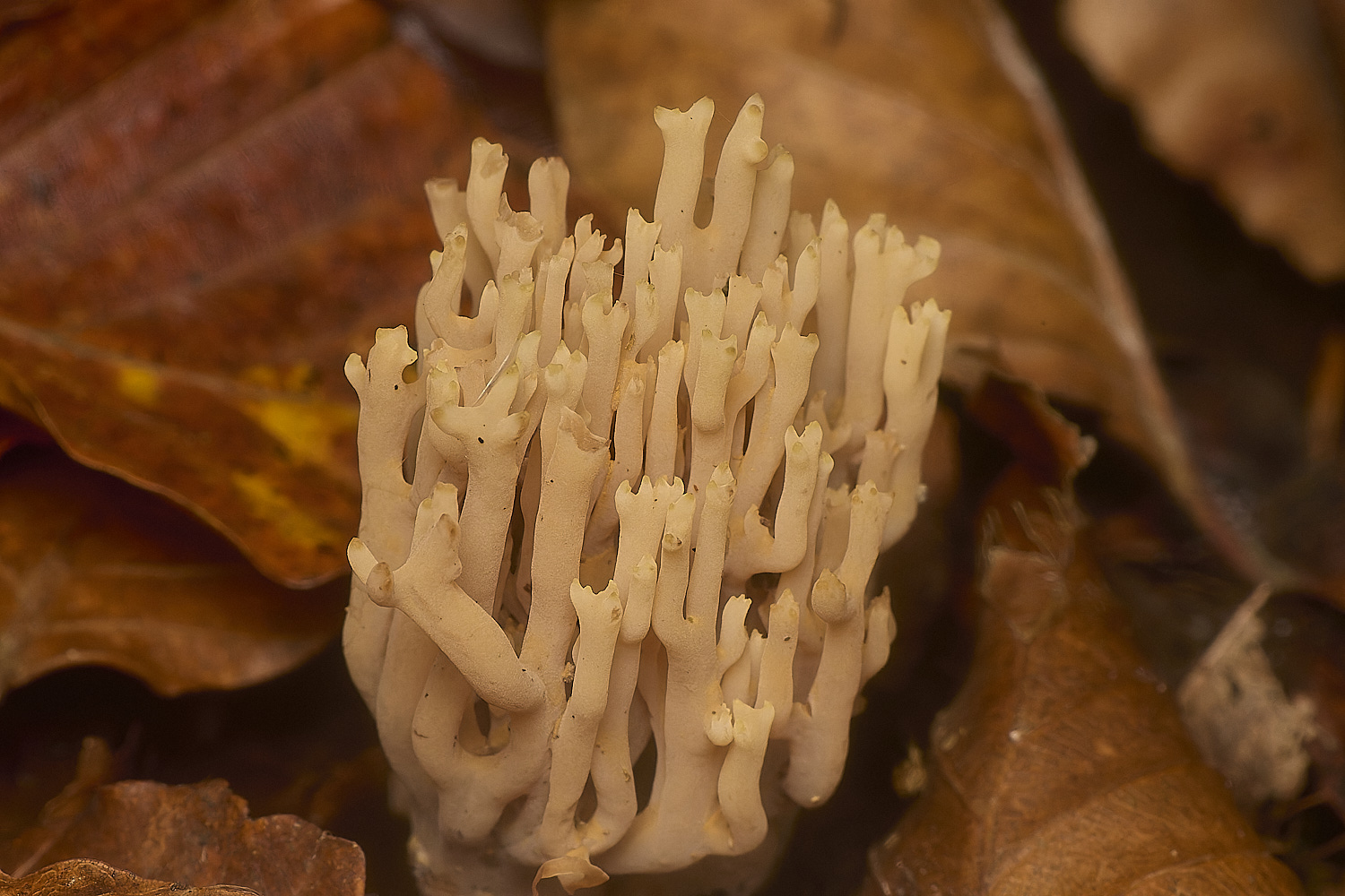
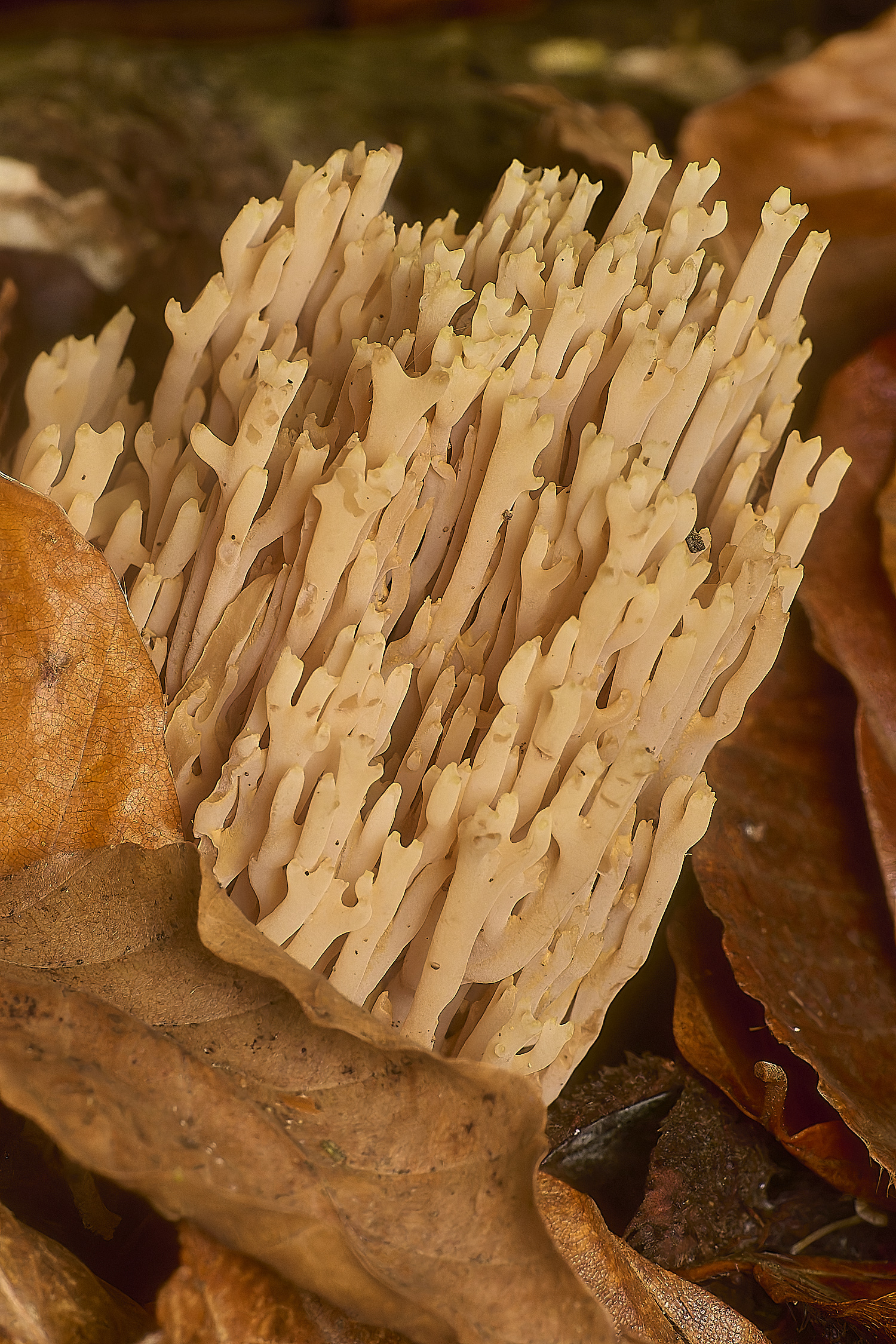
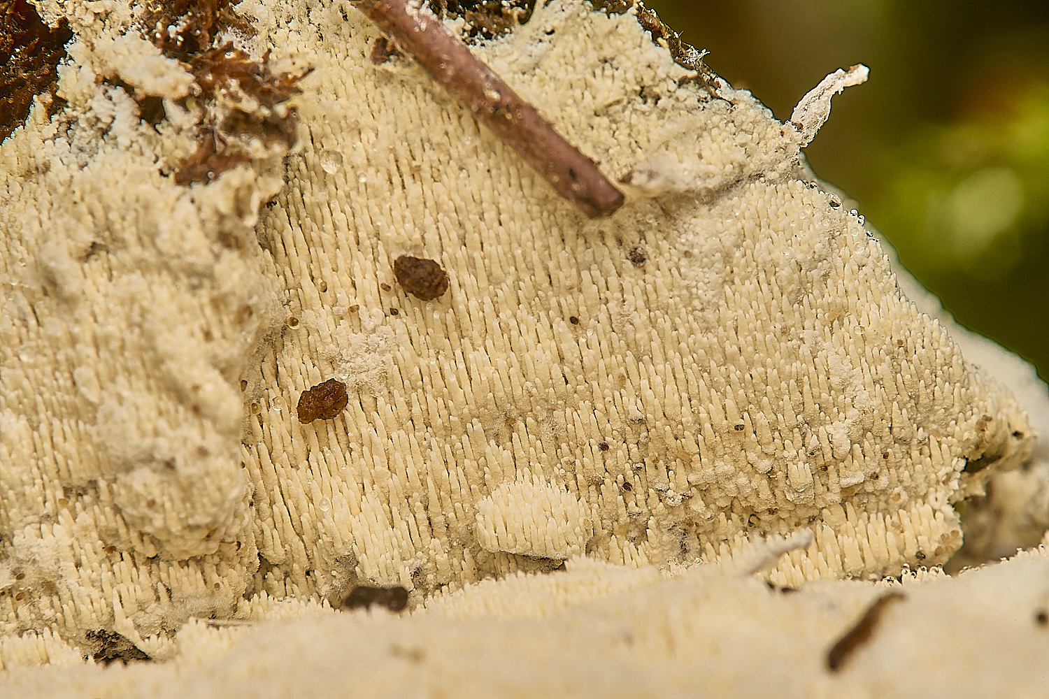
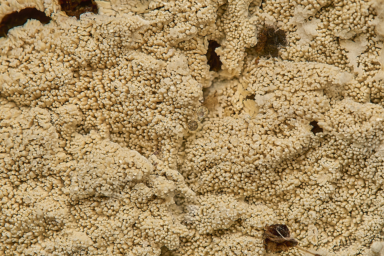
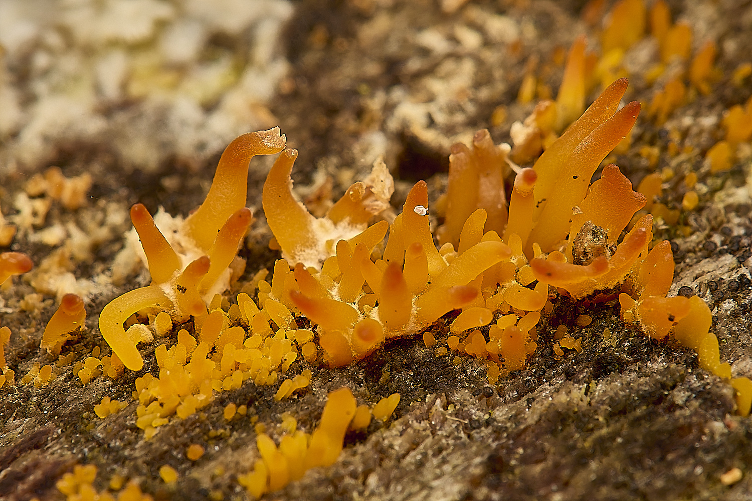
Probably Upright Coral (Ramaria stricta) (Under Beech)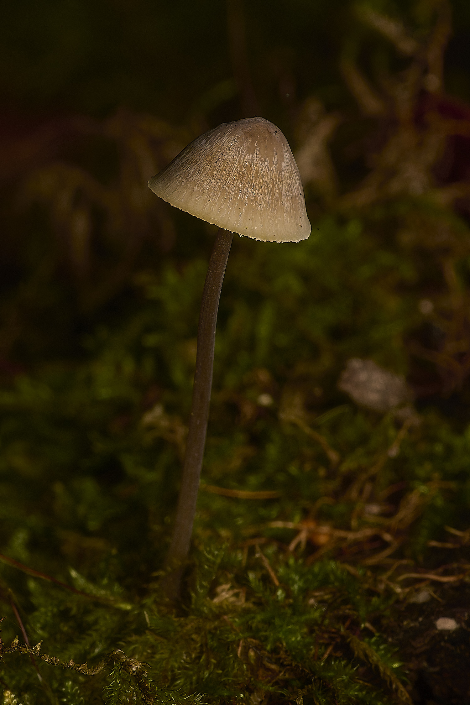
Mycena Sp?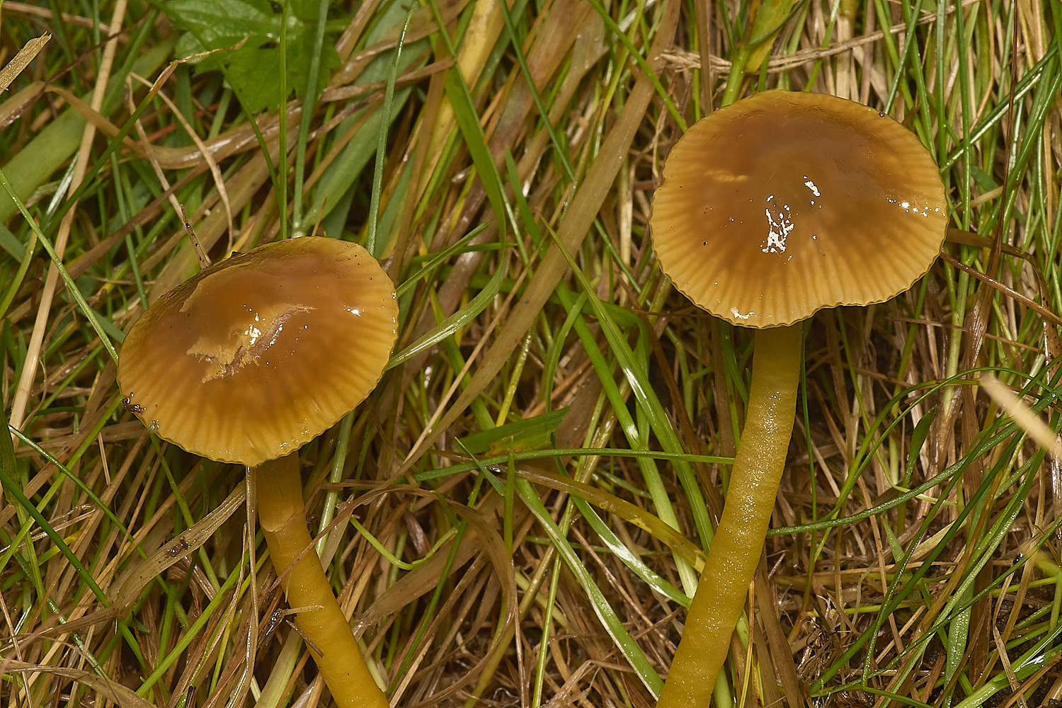
Parrot Waxcap (Gliophurus psittacinus)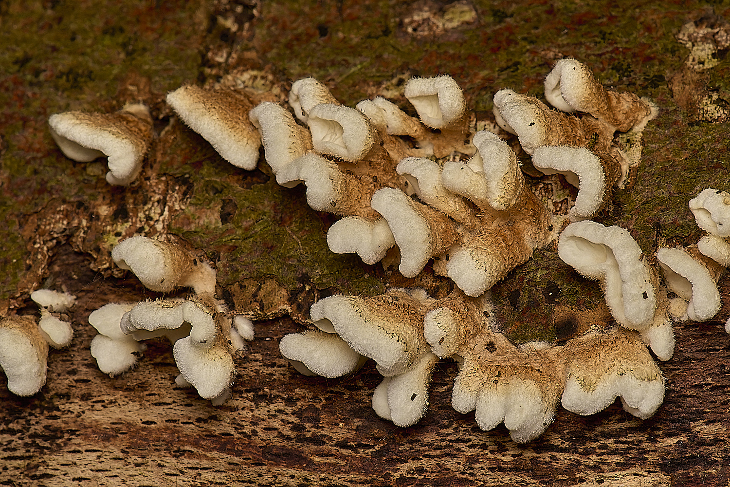
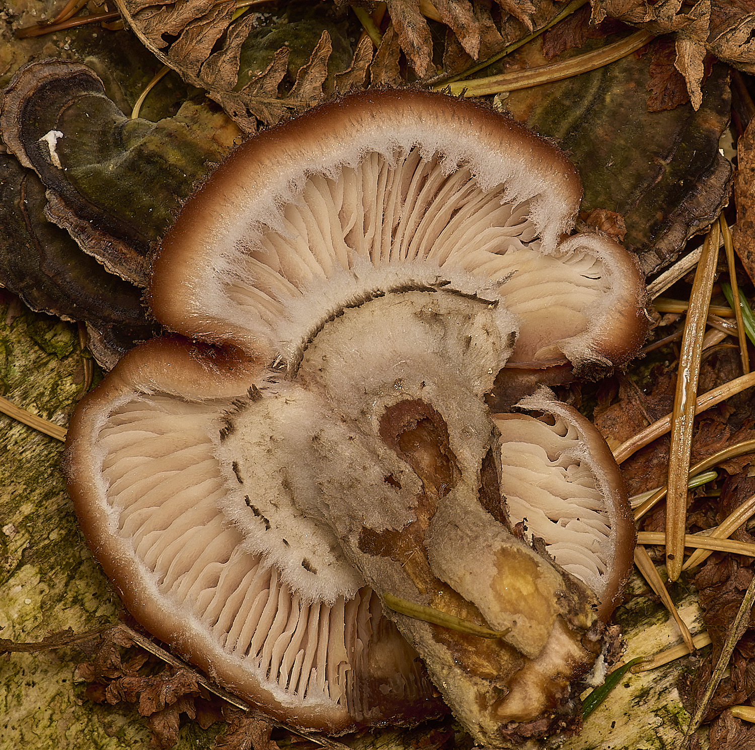
Crimp Gill (Plicatura crispa)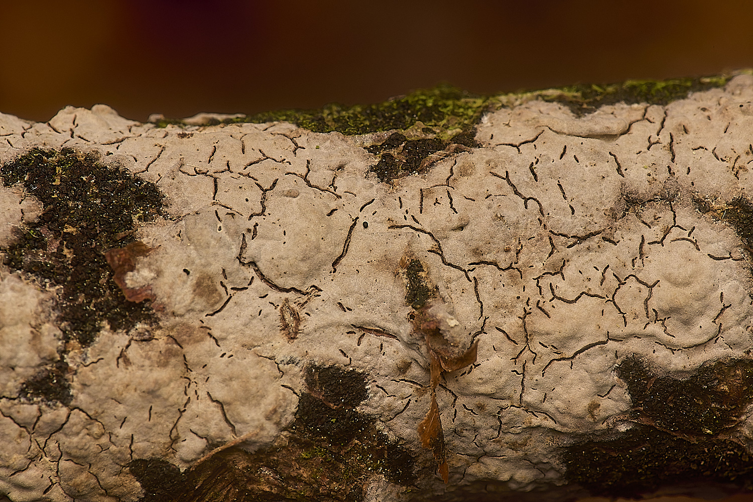
Peniophora Sp?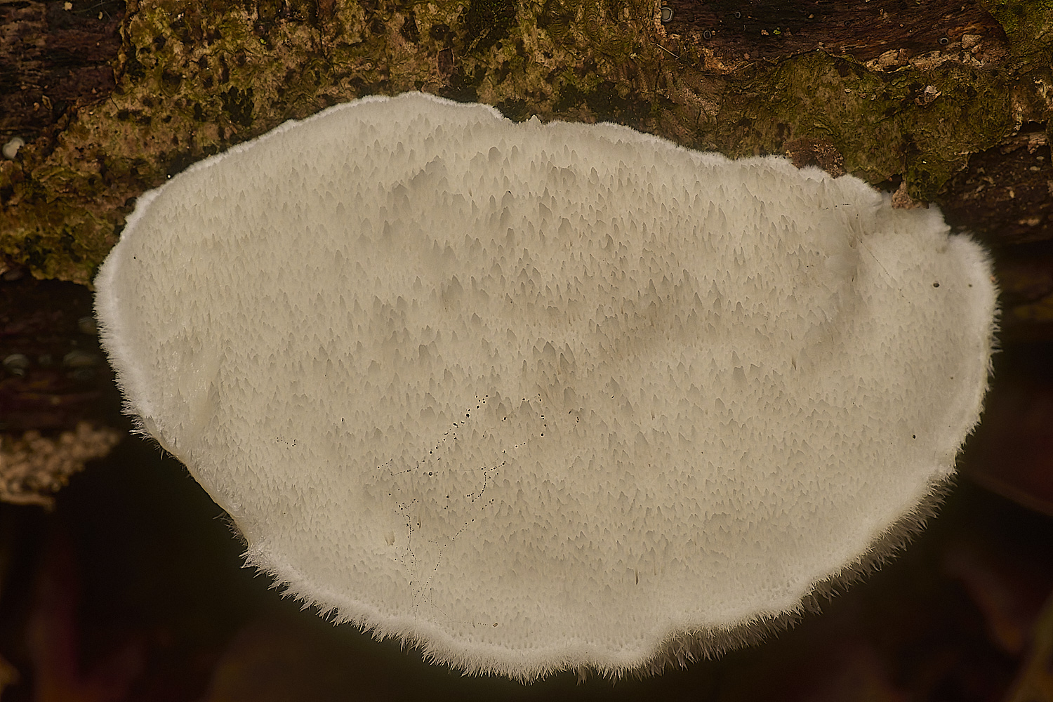
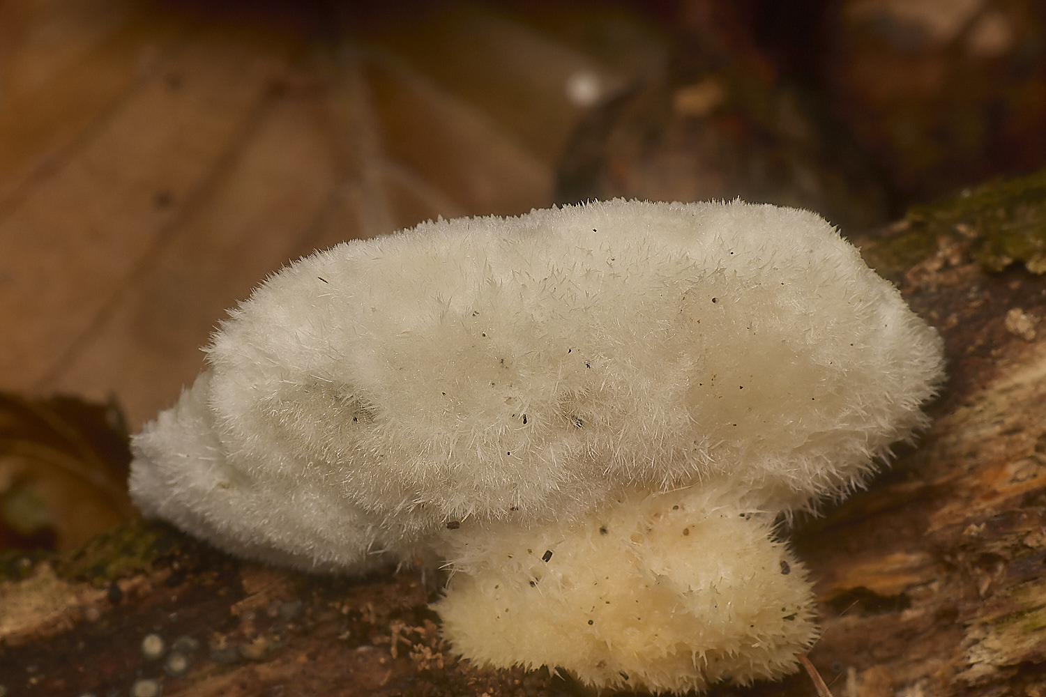
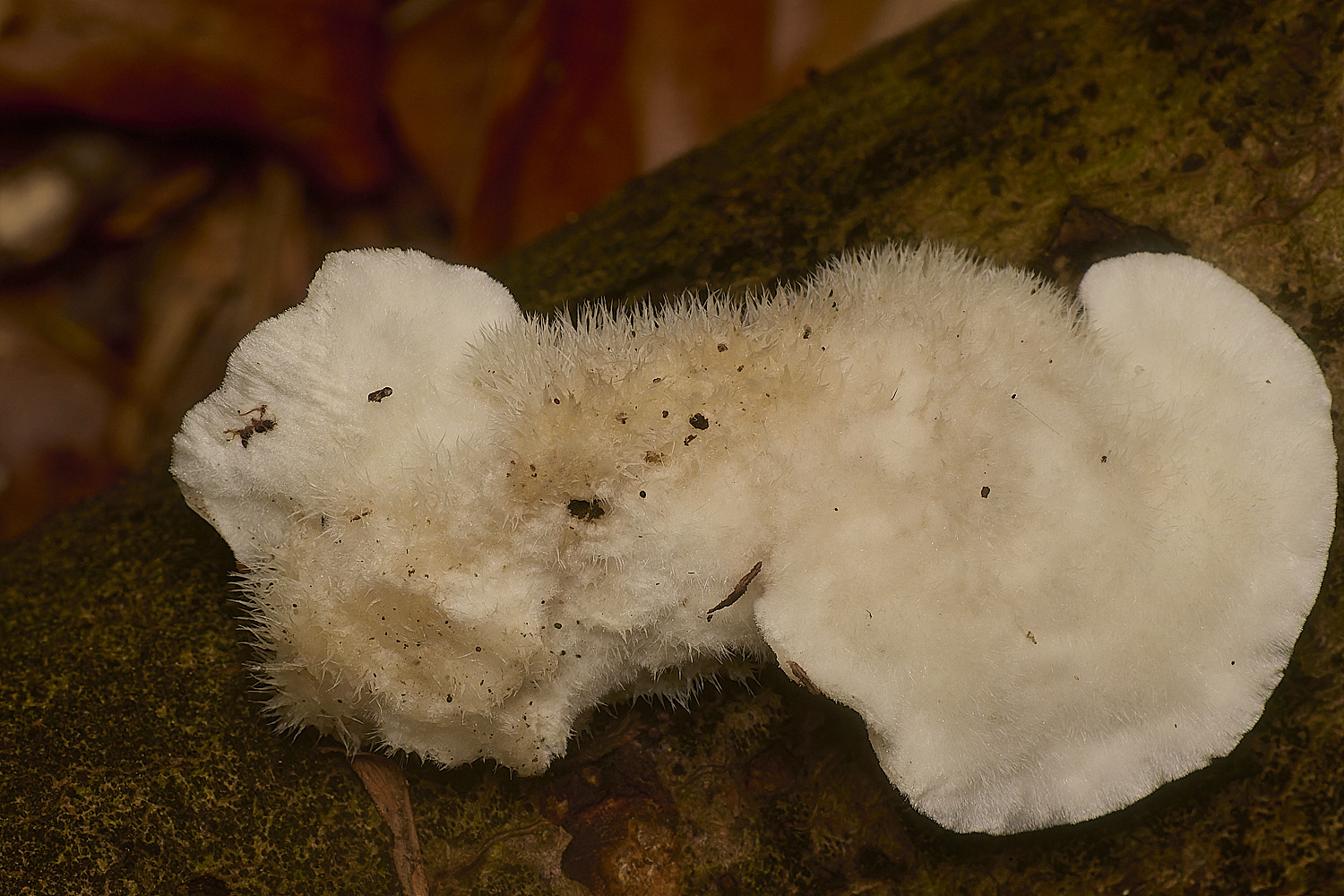
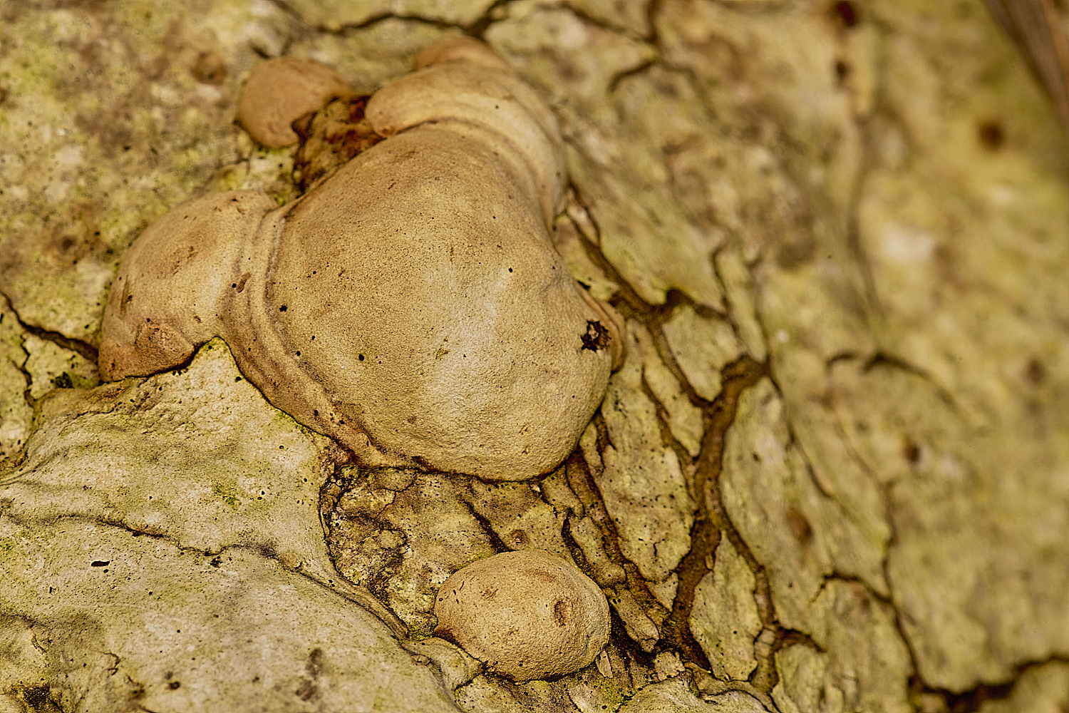
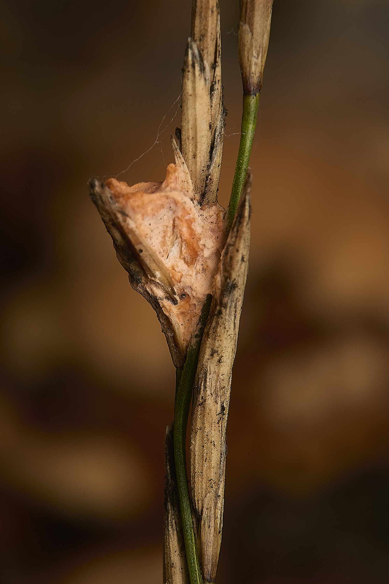
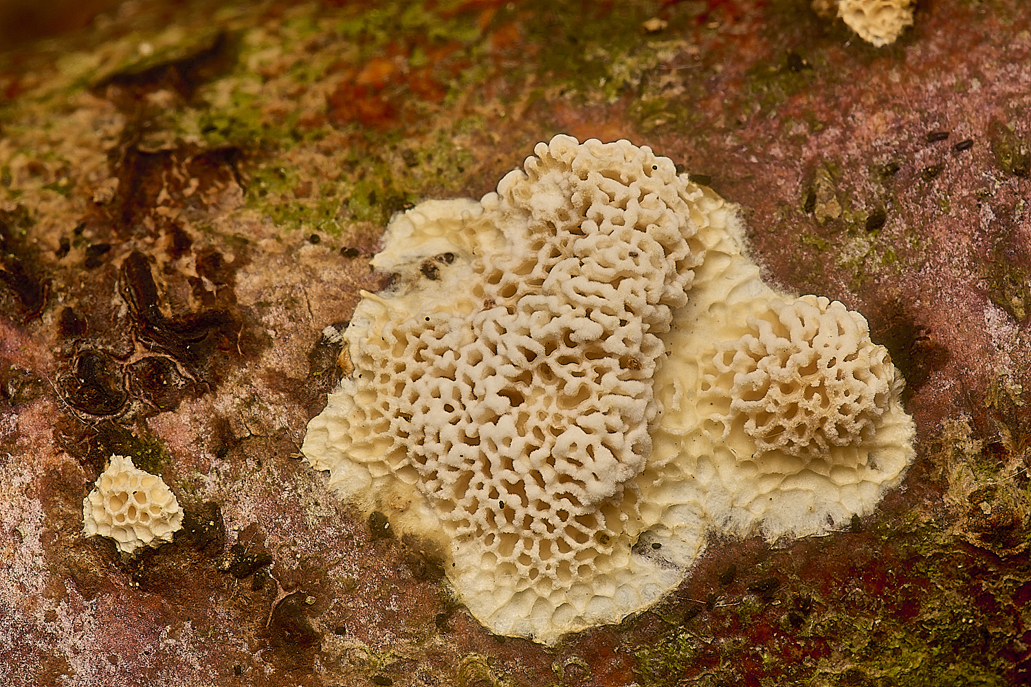
Powderpuff Bracket (Postia ptychogaster)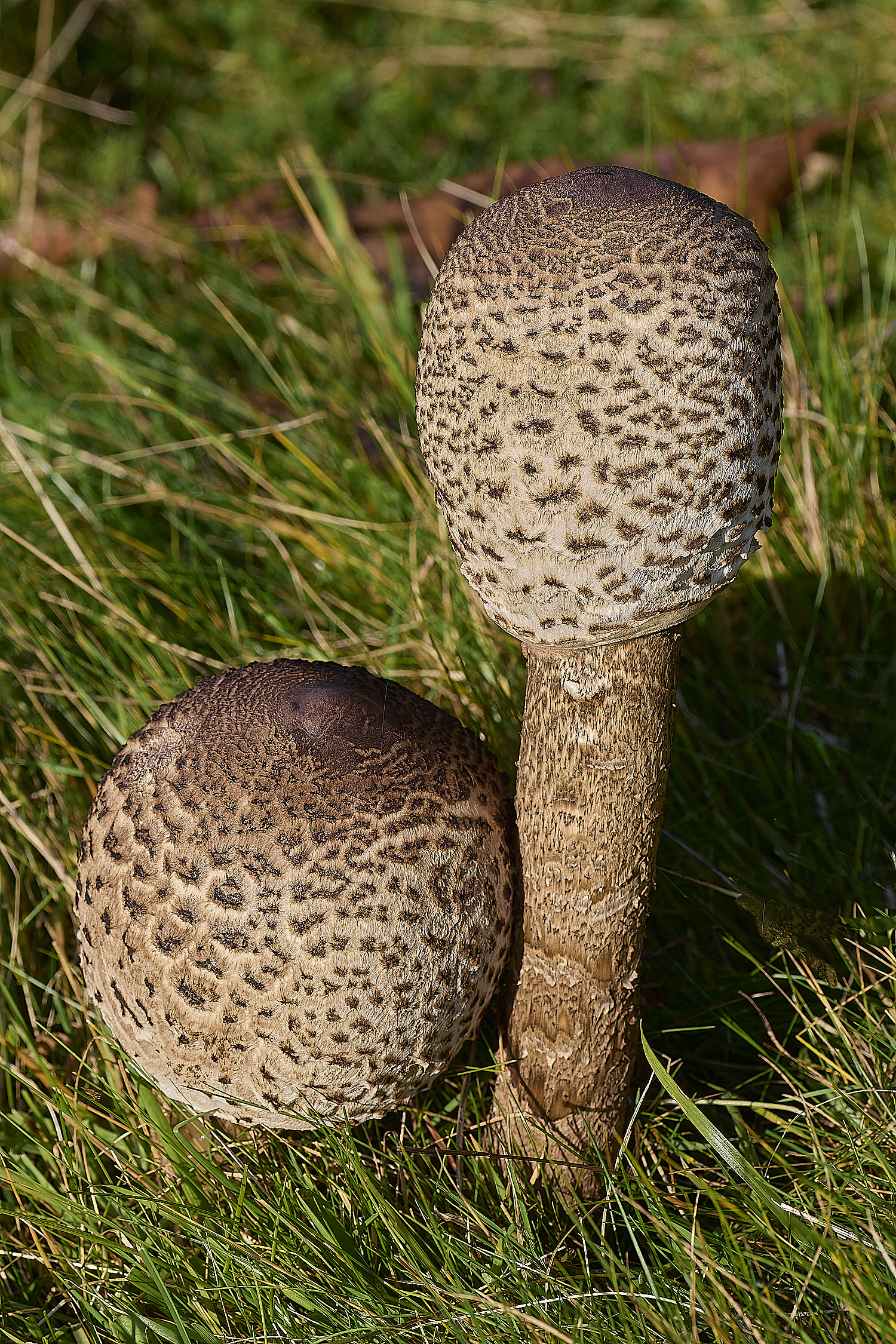
Shaggy Parasol Chlorophyllum rhacodes)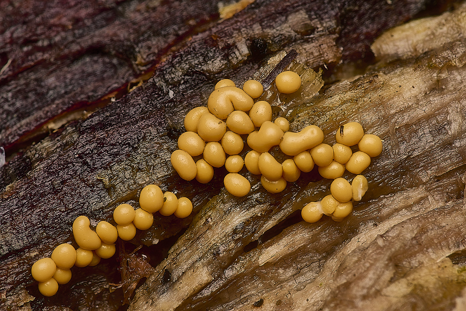
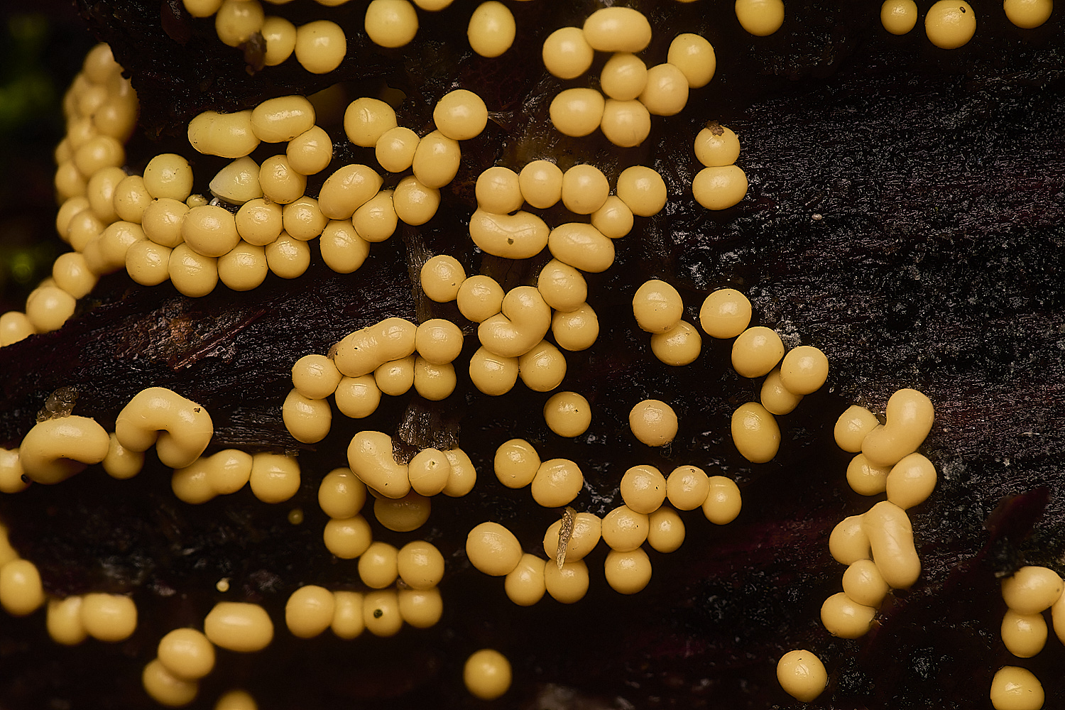
Slime Mold Sp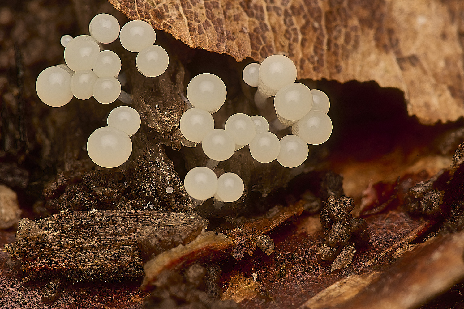
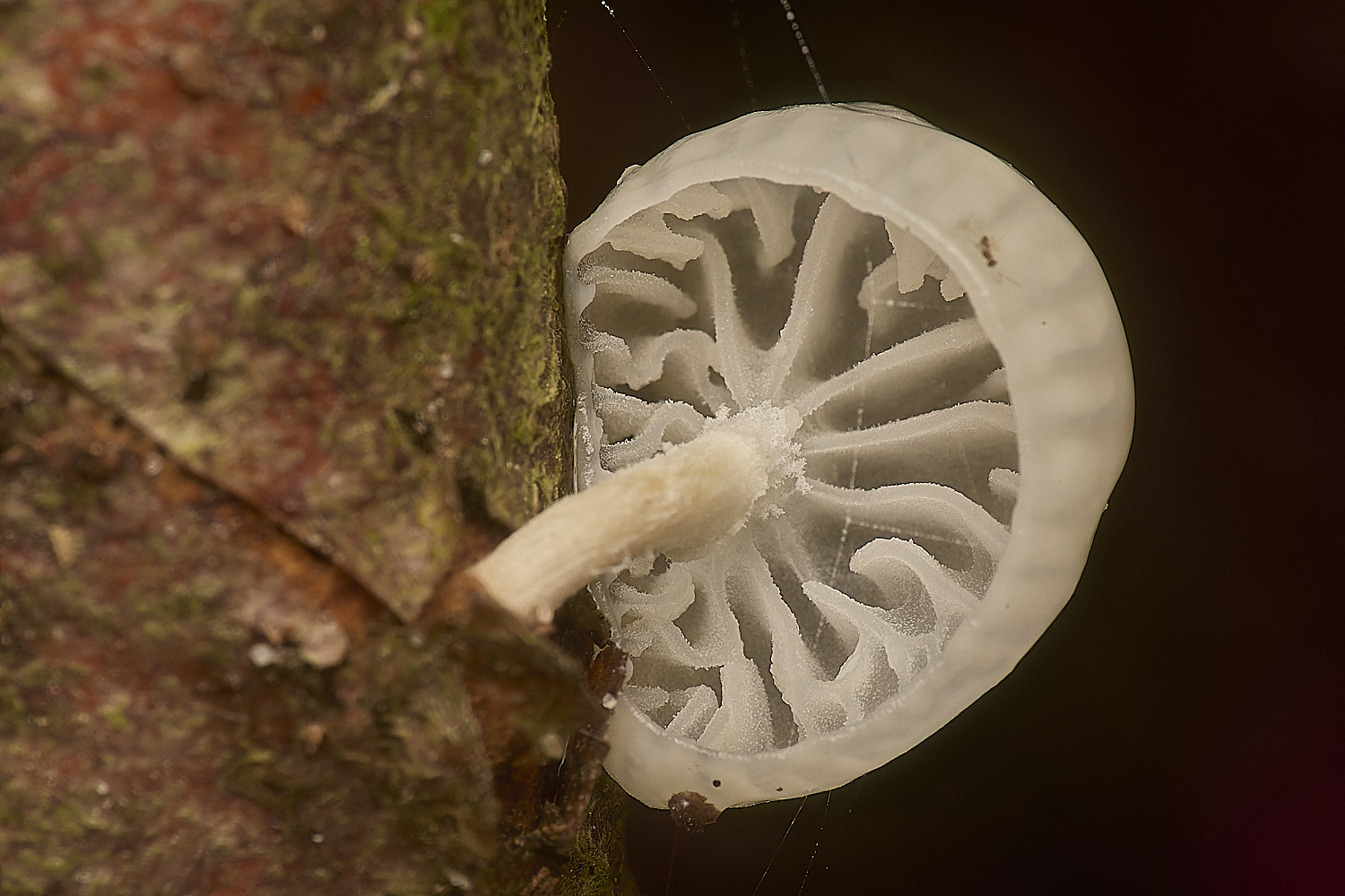
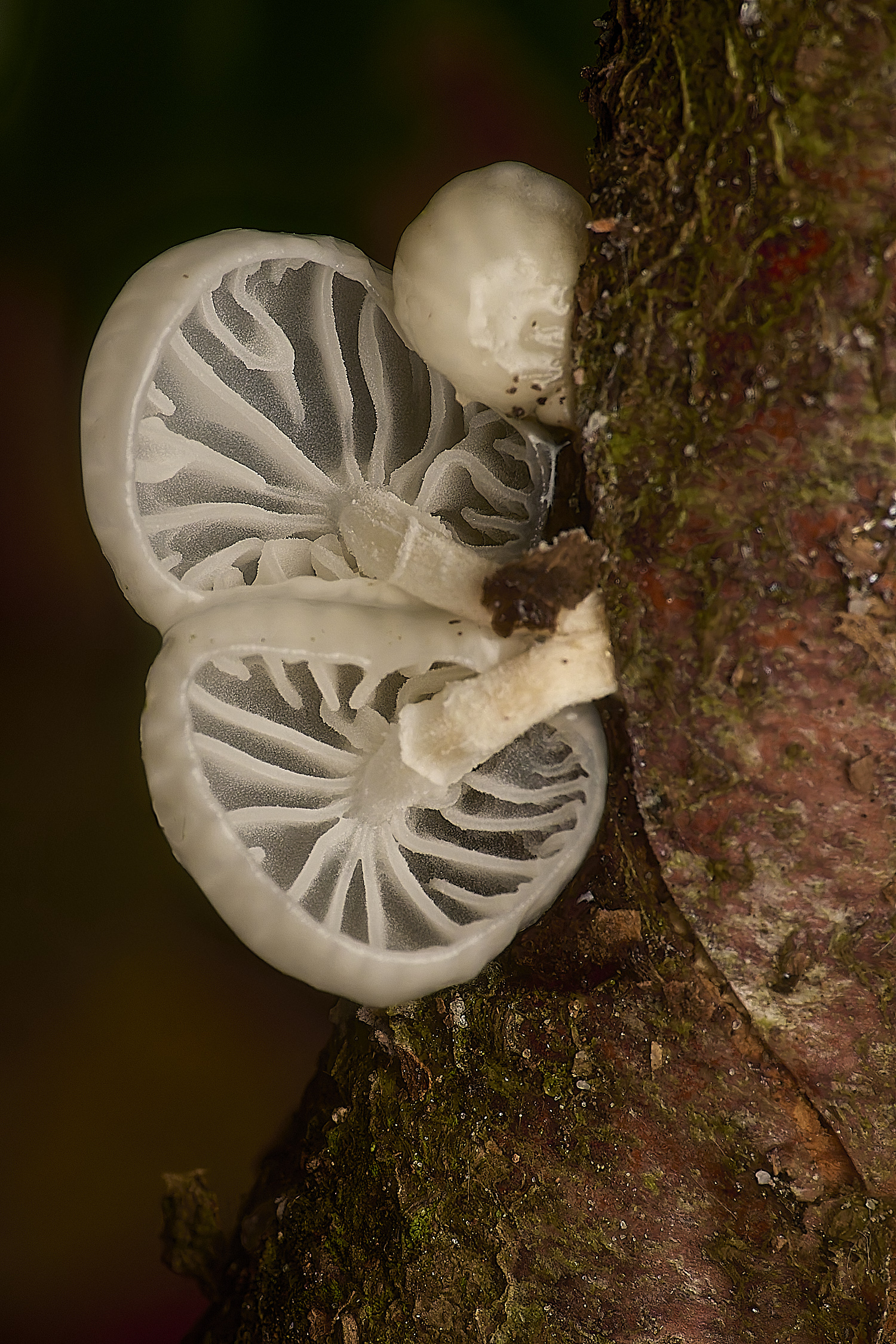
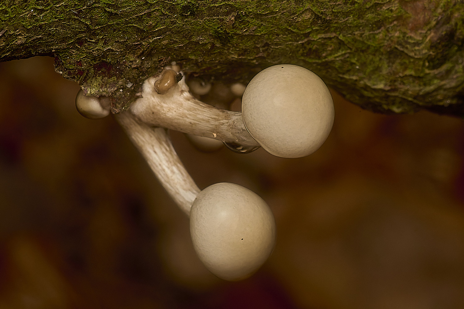
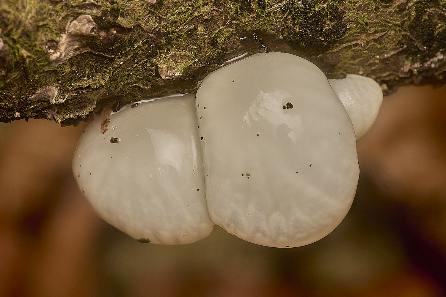
Porcelain Fungus (Oudemansiella mucida)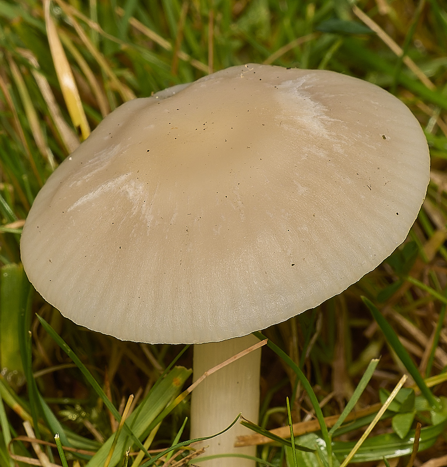
Snowy Waxcap (Cupophyllus virgineus)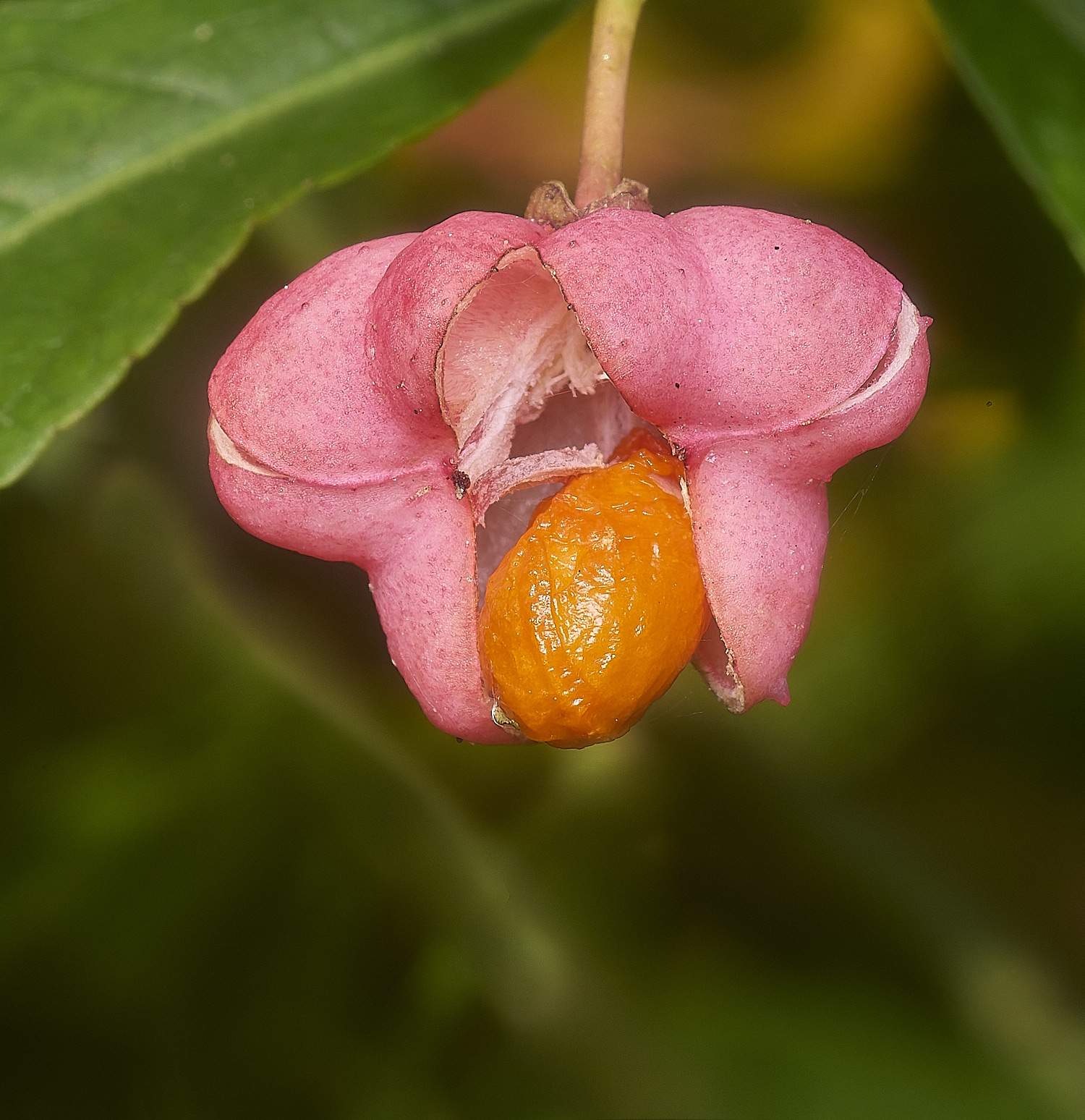
Spindle berry (Euonymus europaeus)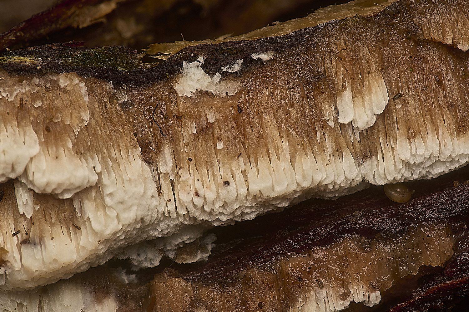
?
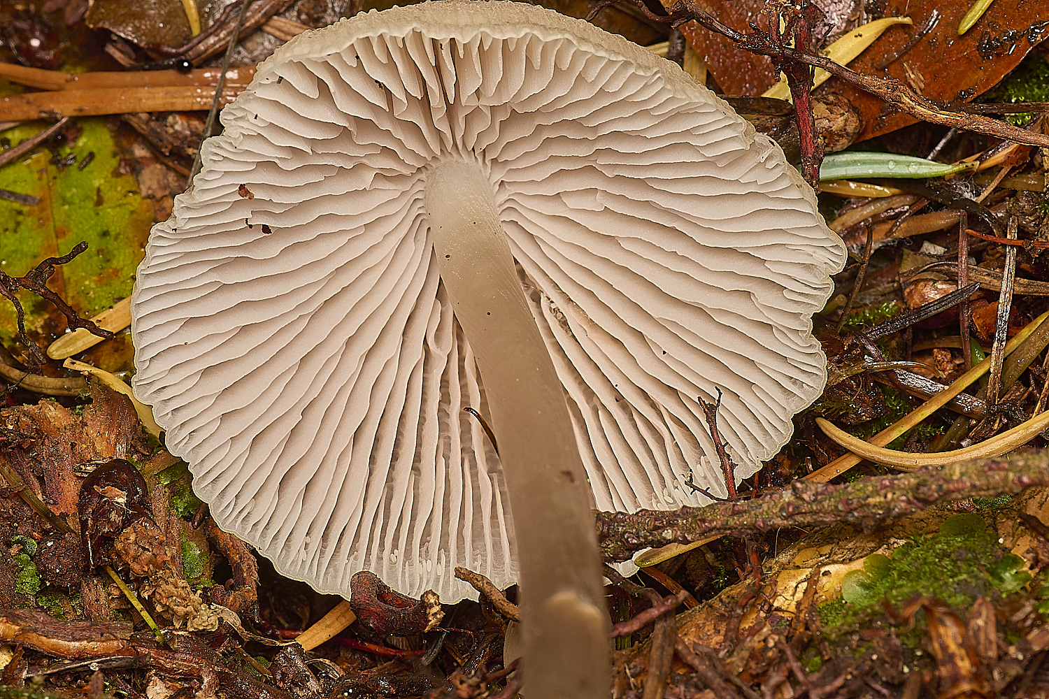
Toughshank Sp?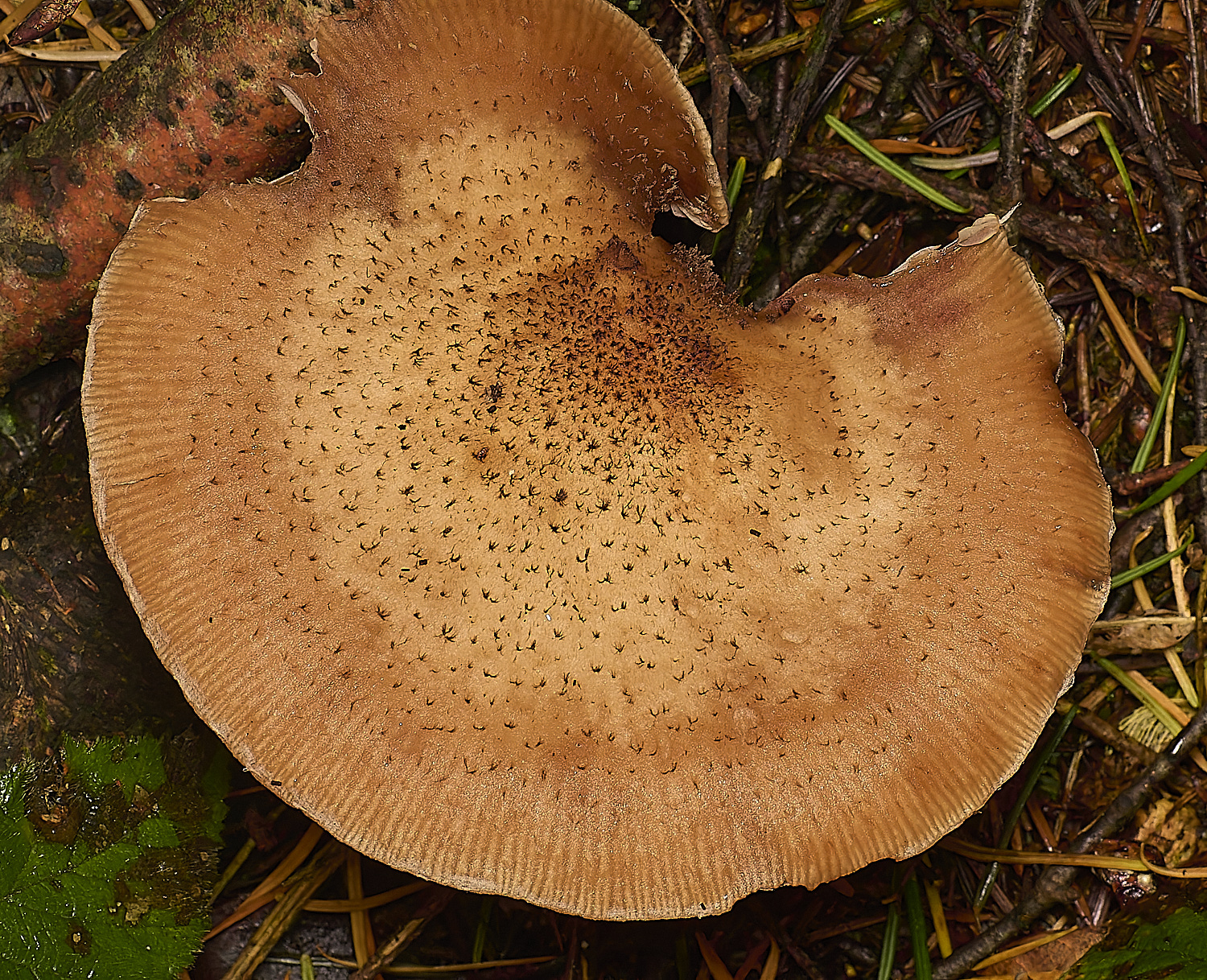
Honey Fungus (Armillaria mellea)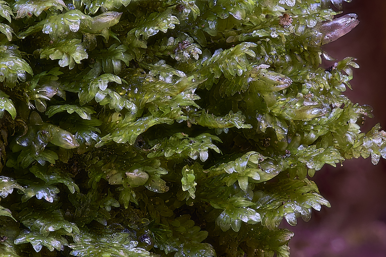
White Earwort (Diplophyllum albicans)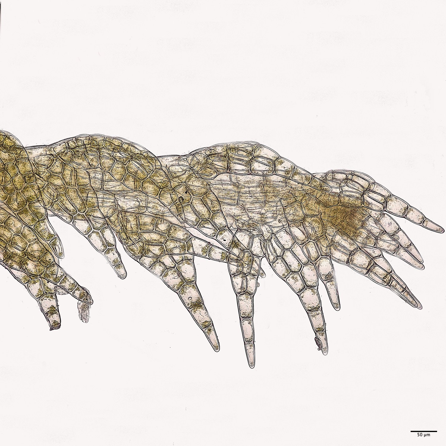
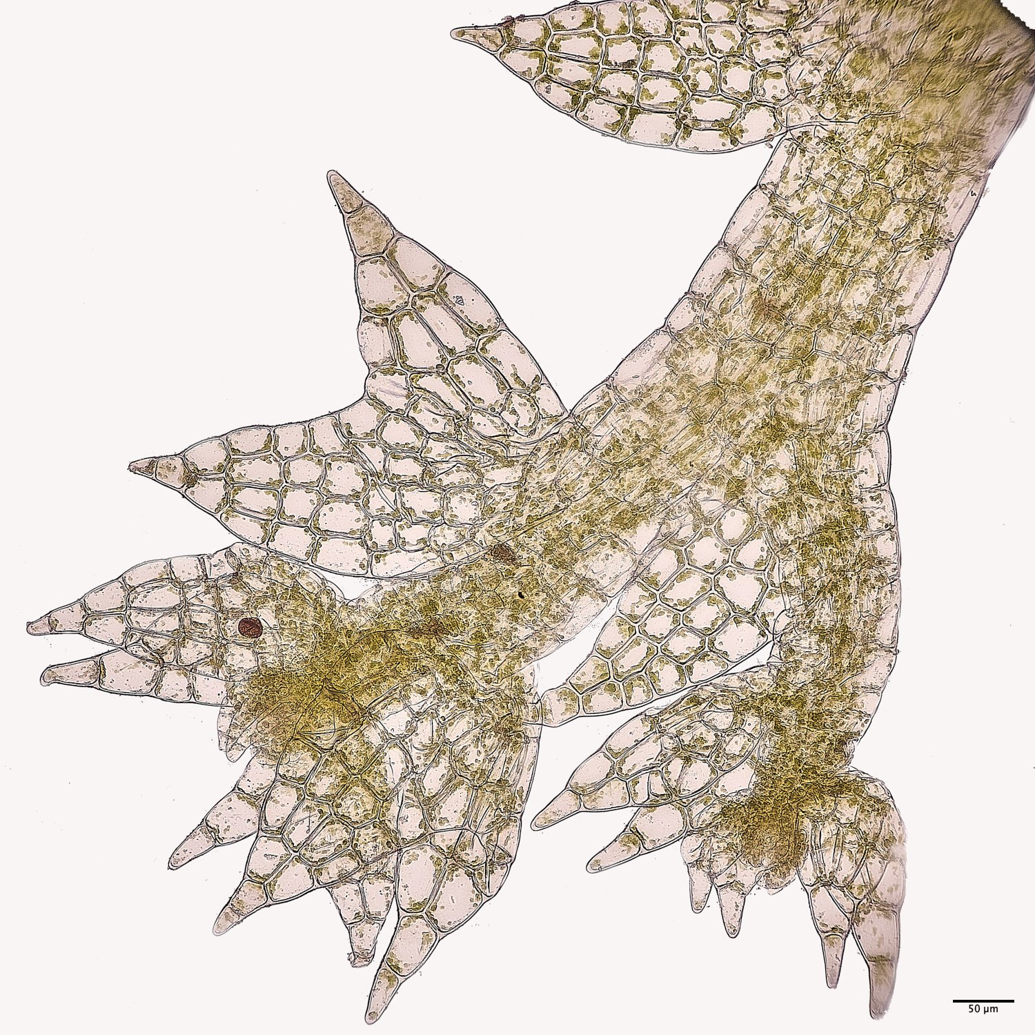
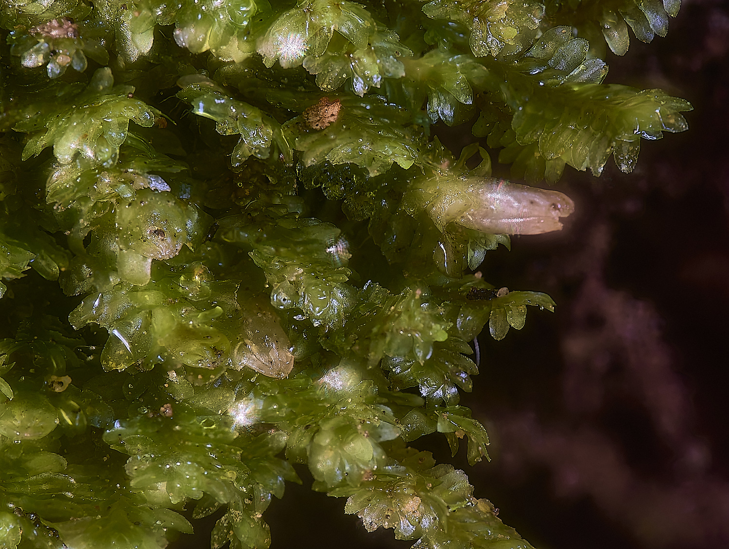
Forcipated Pincerwort (Cephalozia connivens)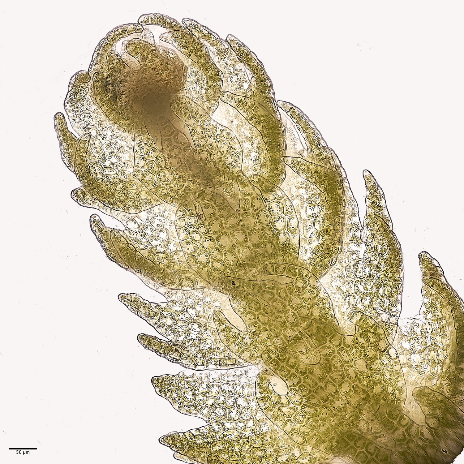
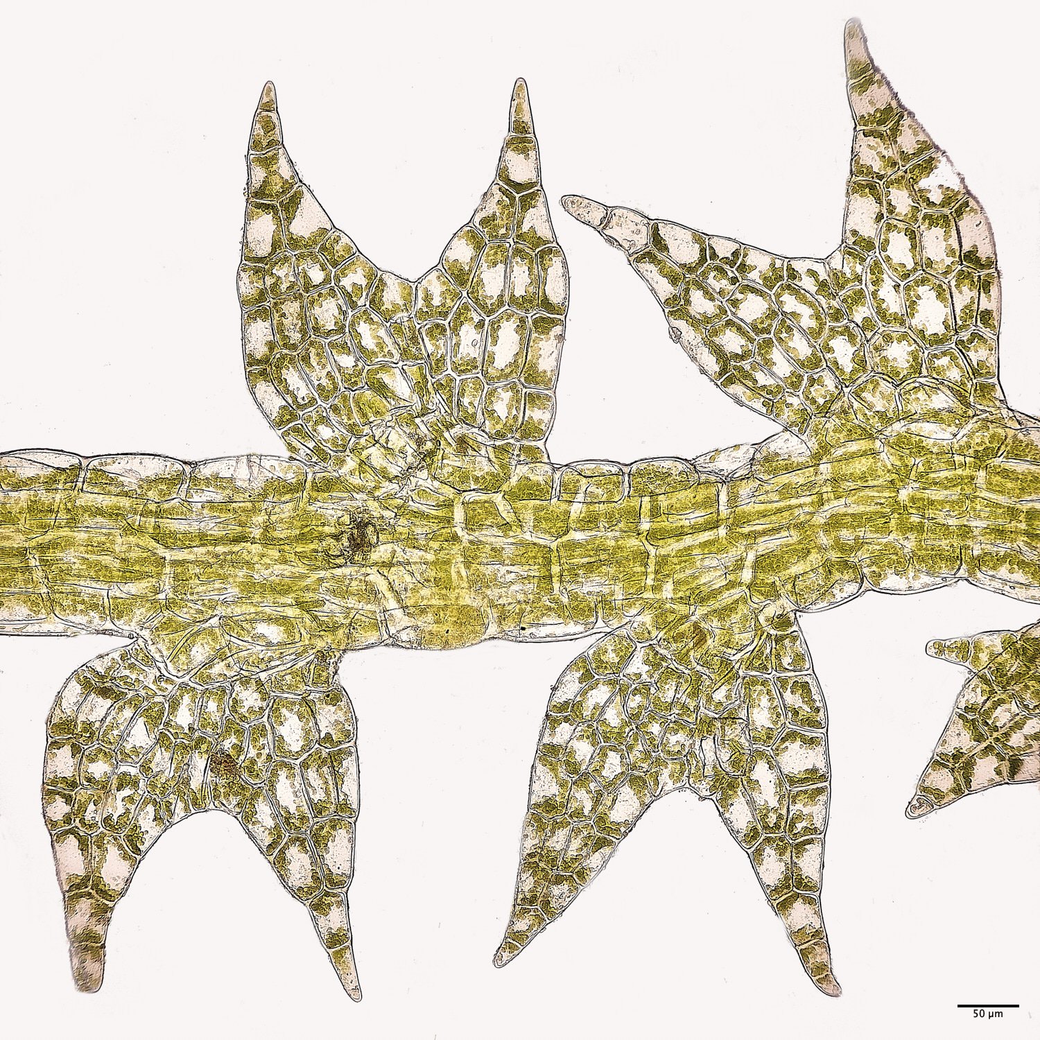
Creeping Fingerwort (Lepidozia reptans)