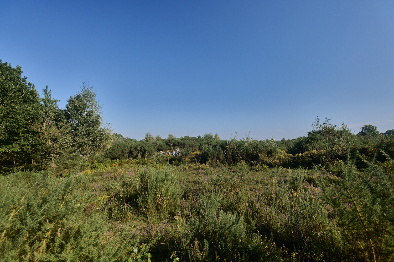
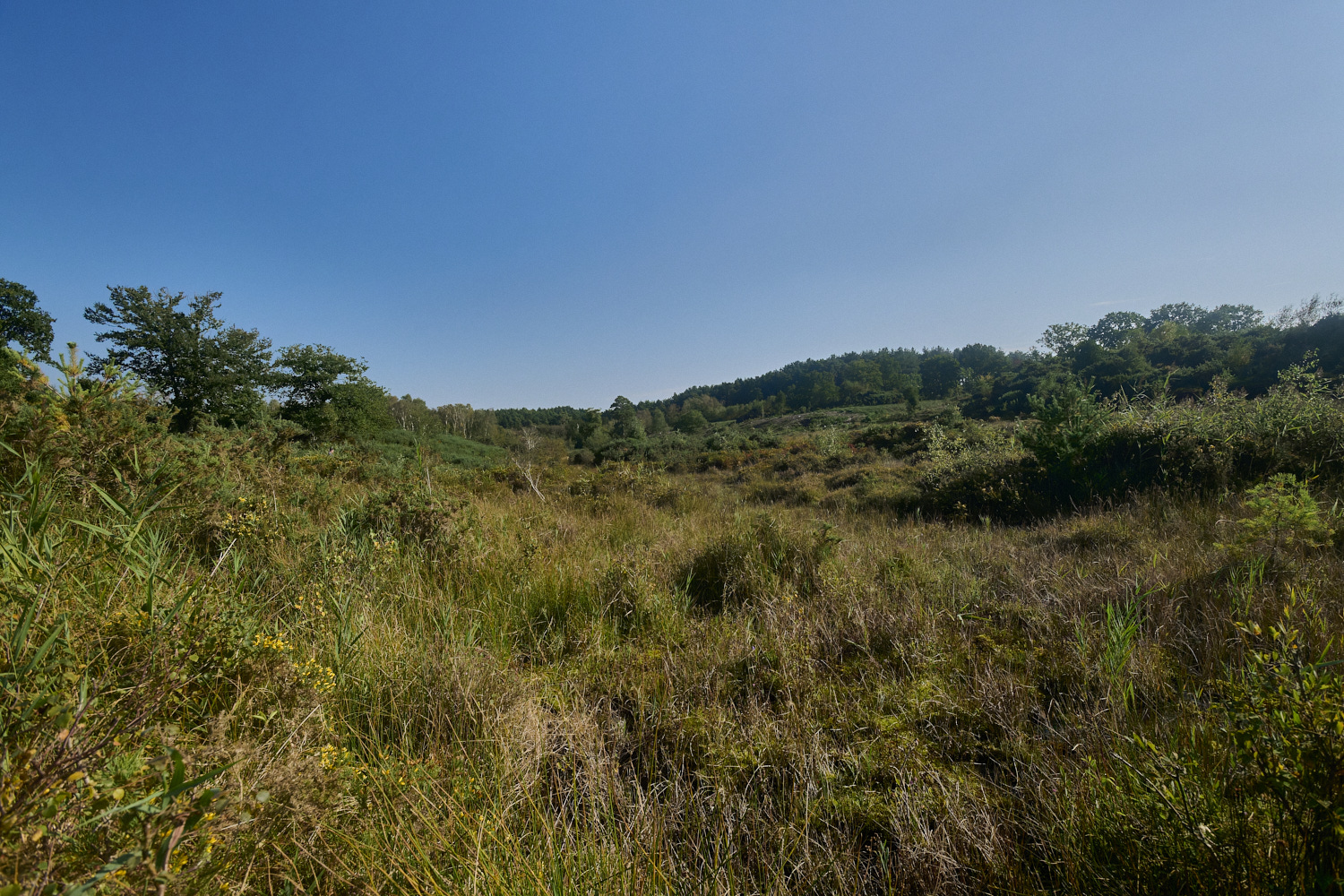
Holt Lowes
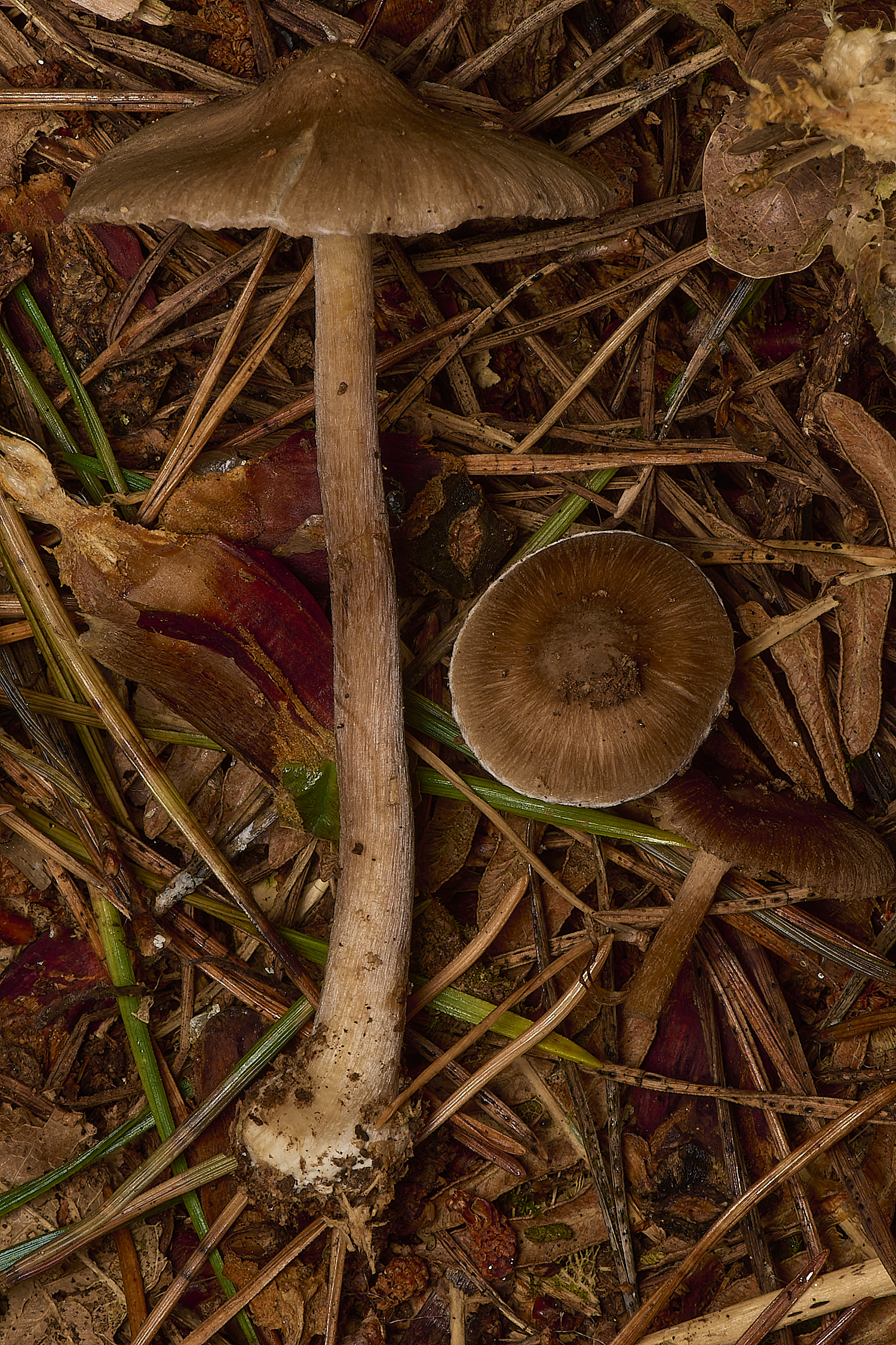
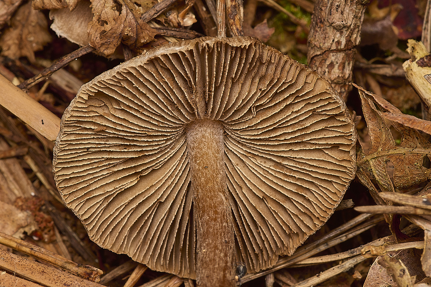
Incoybe napipes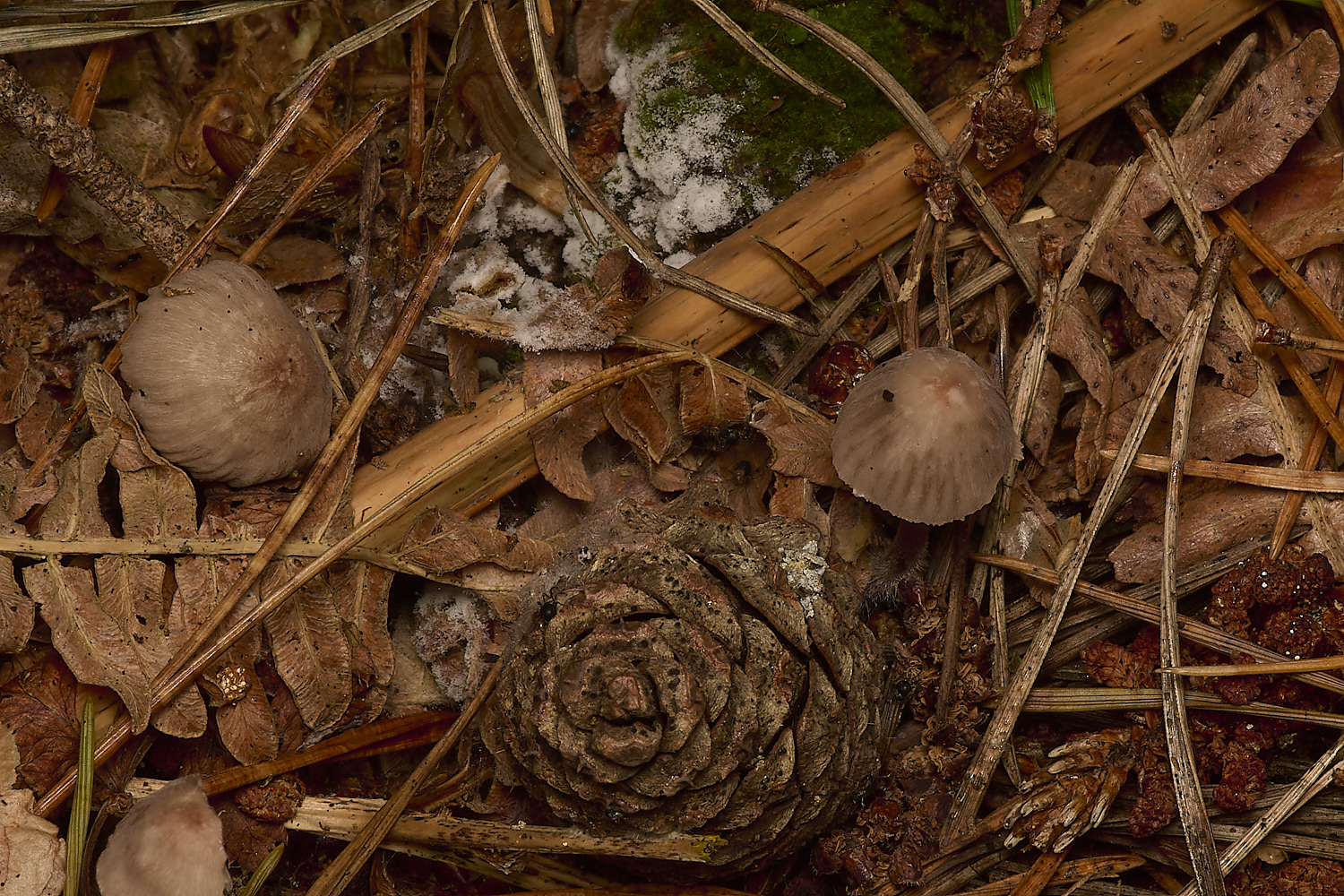
Mycena capillaripes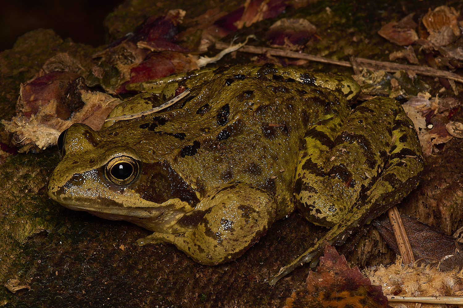
Frog (Rana temproraria)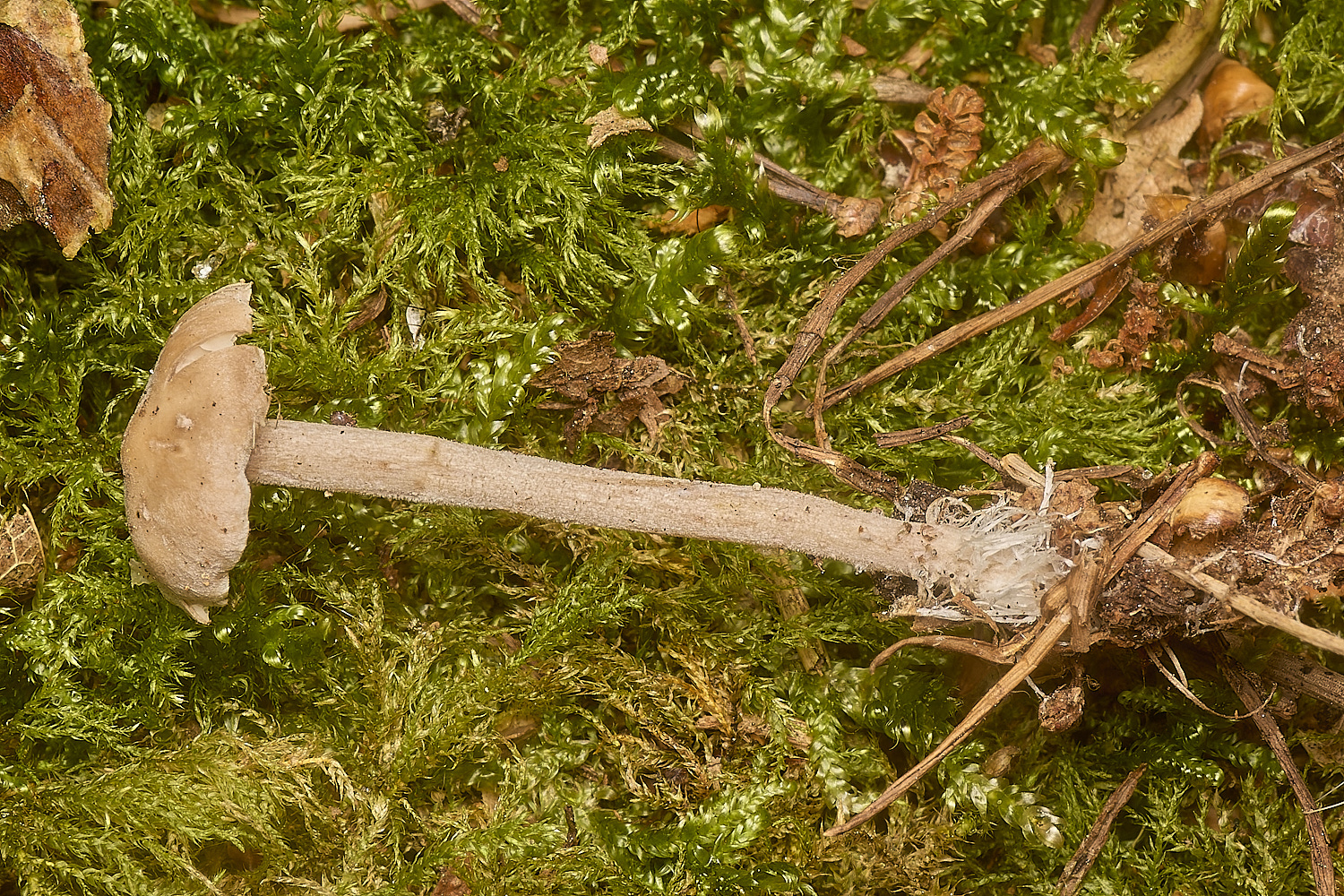
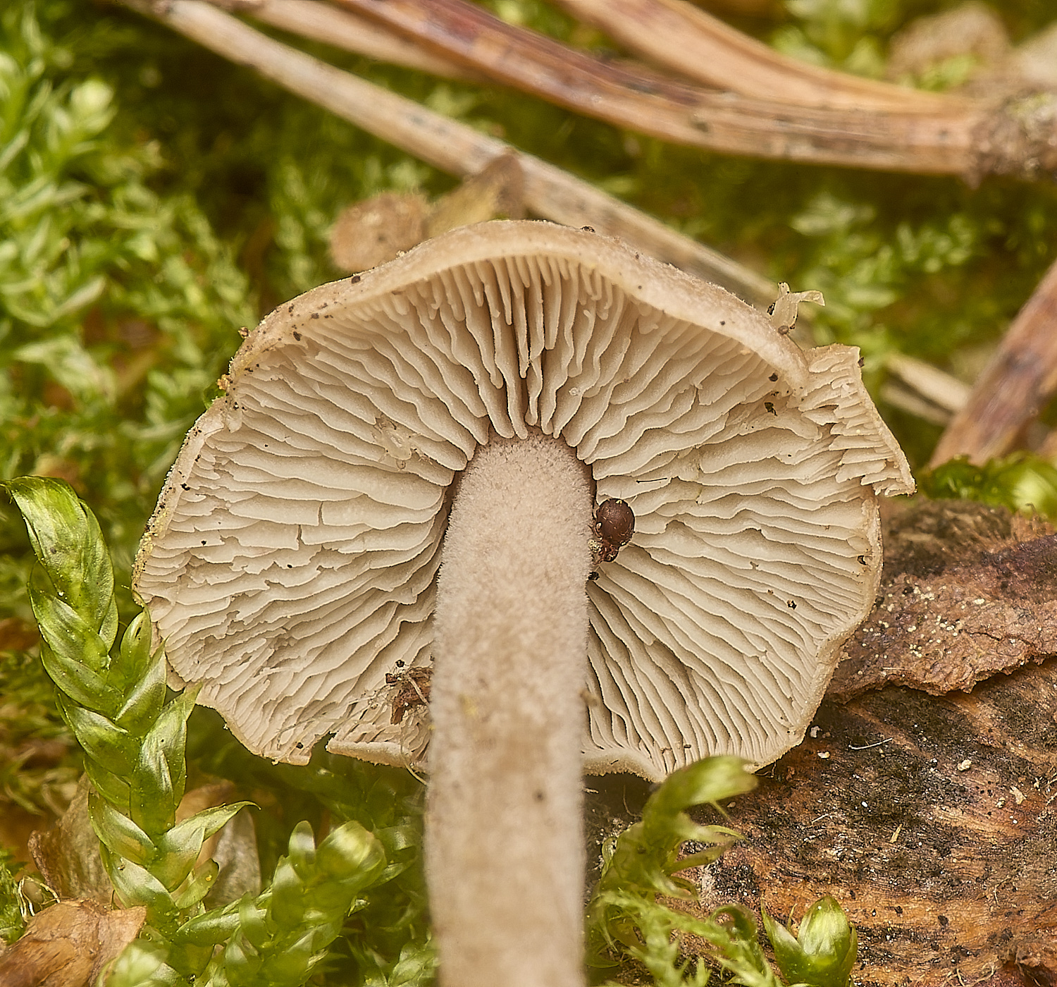
Clustered Toughshank (Gymnopus confluens)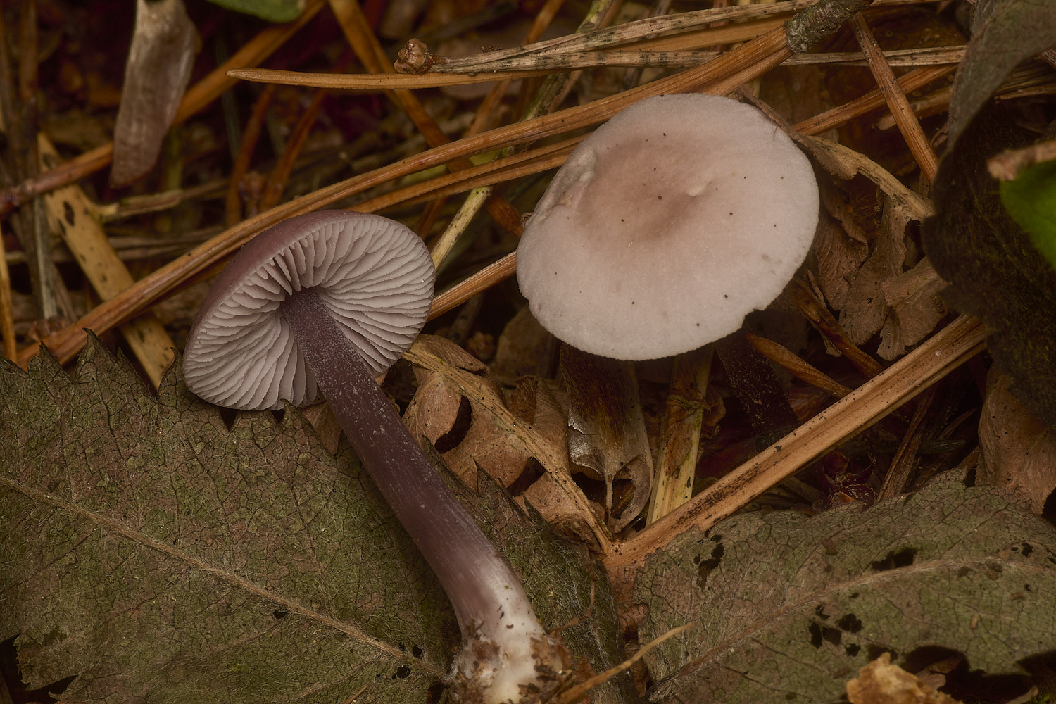
Lilac Bonnet (Mycena pura)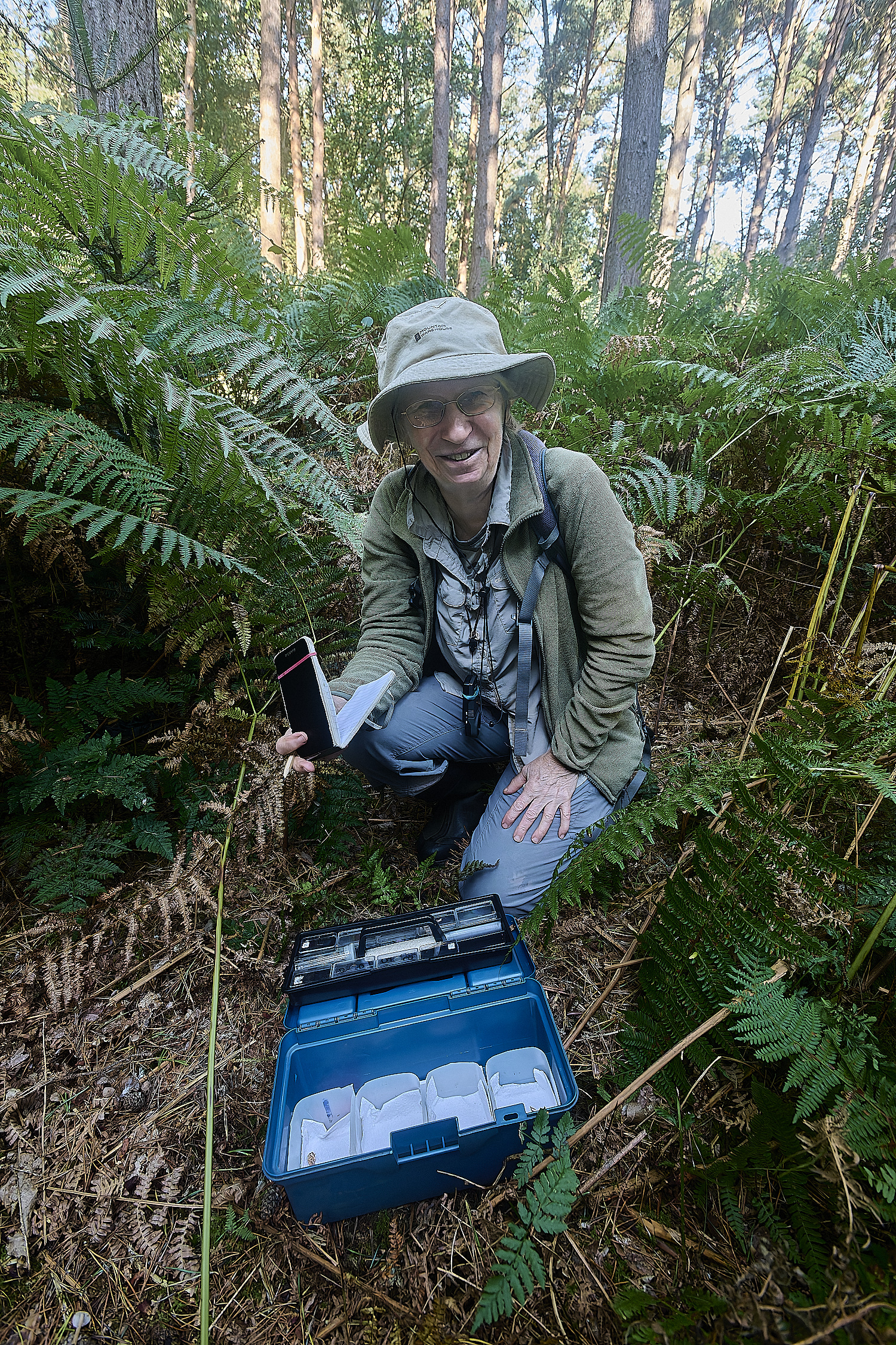
An intrepid mycologist
DNA collecting method - approved!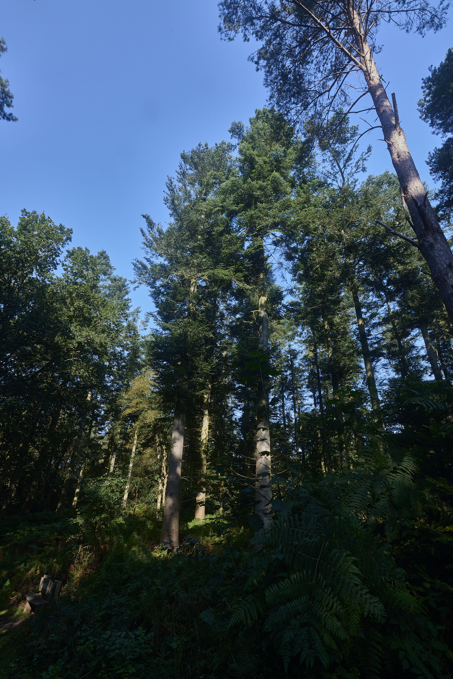
Grand Fir (Abies grandis)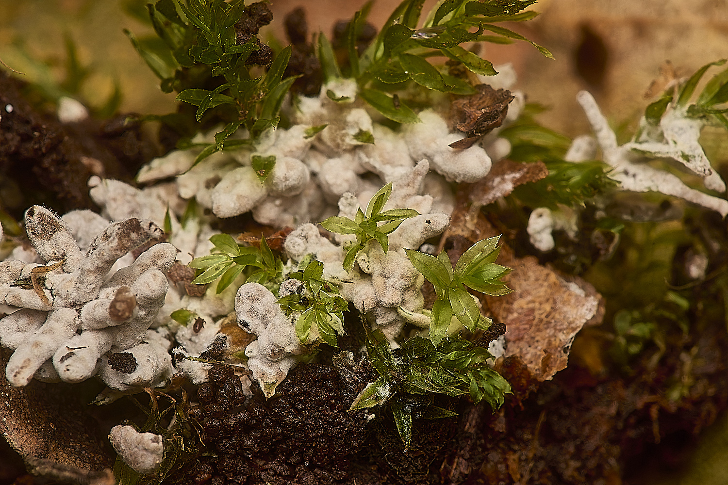
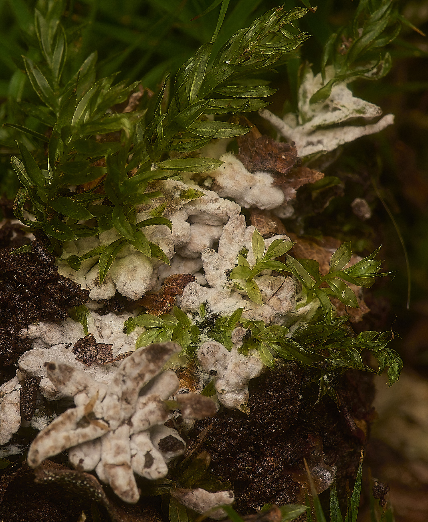
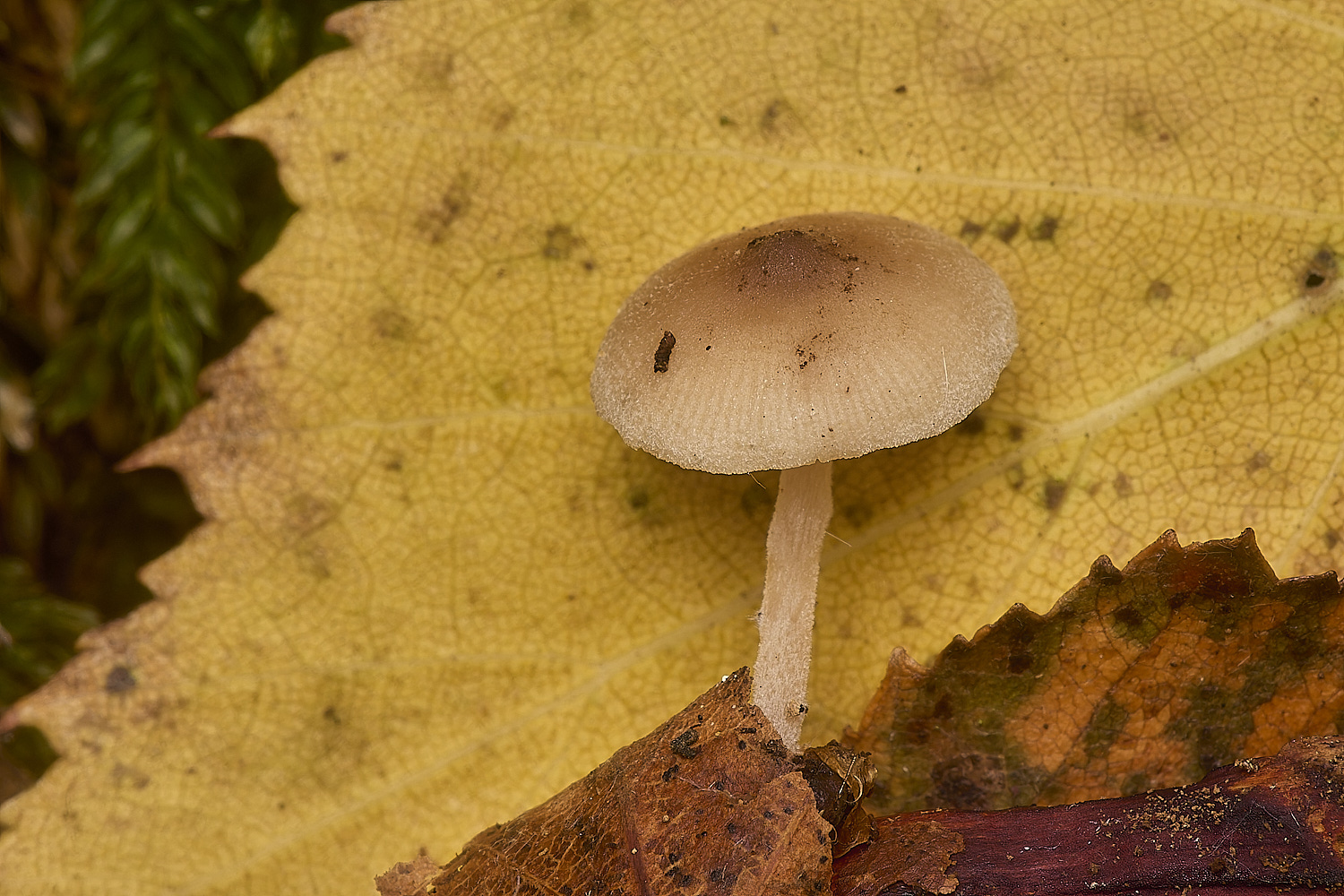
Fungus associated with Swan's-neck Thyme Moss (Mnium hornum)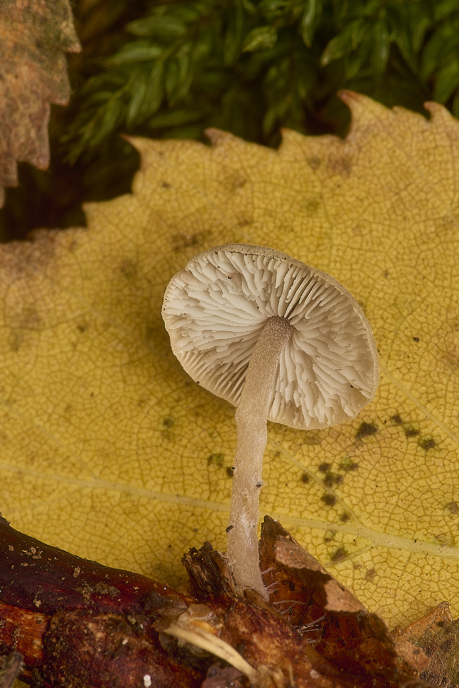
Unknown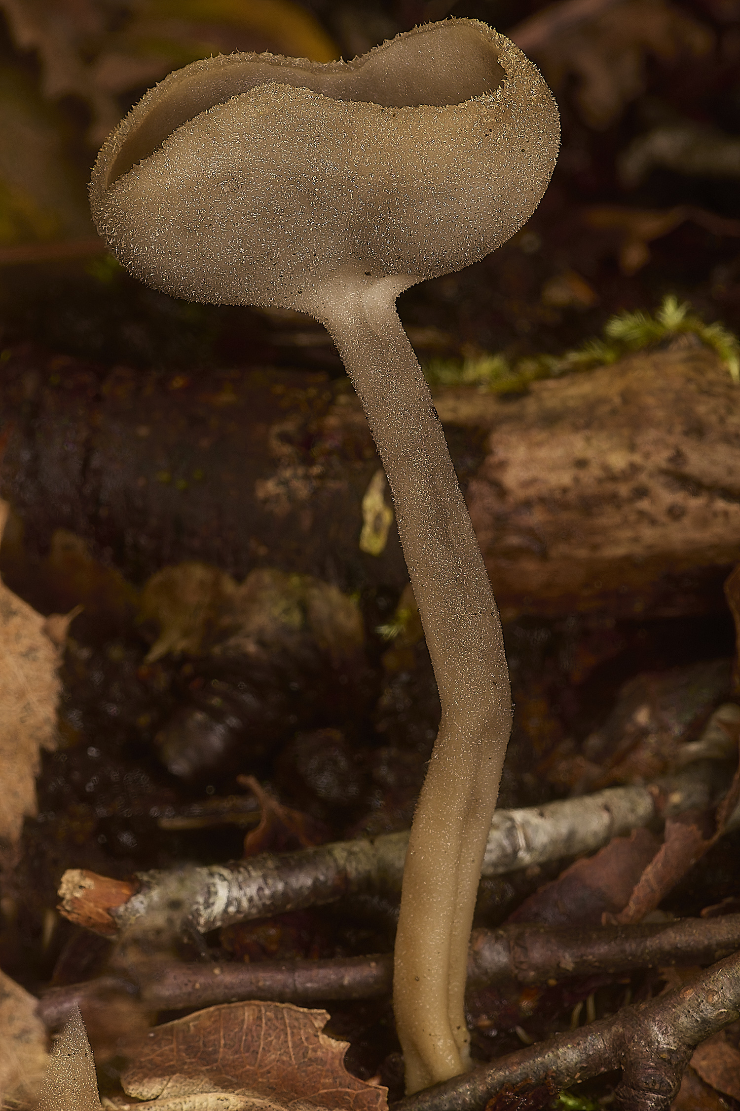
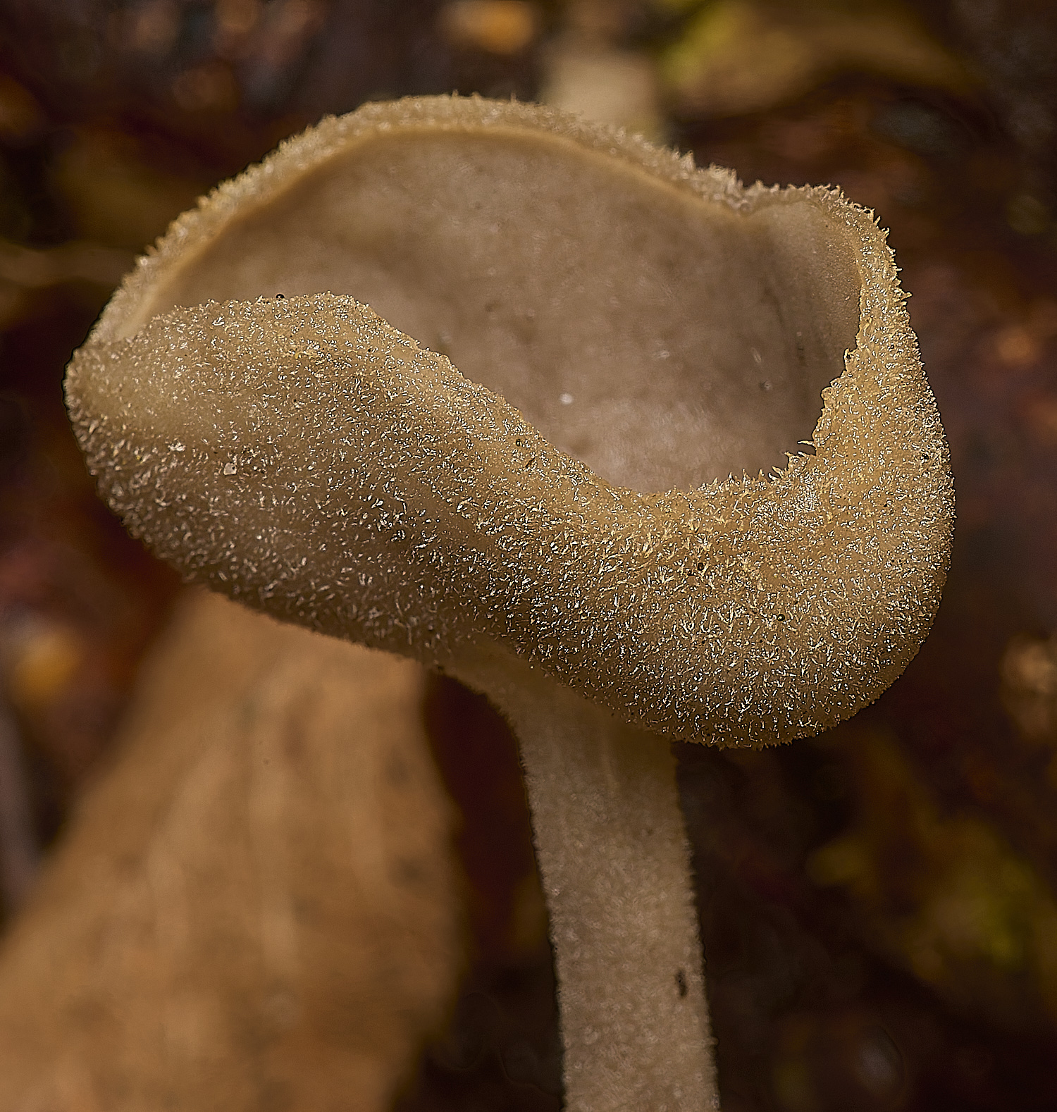
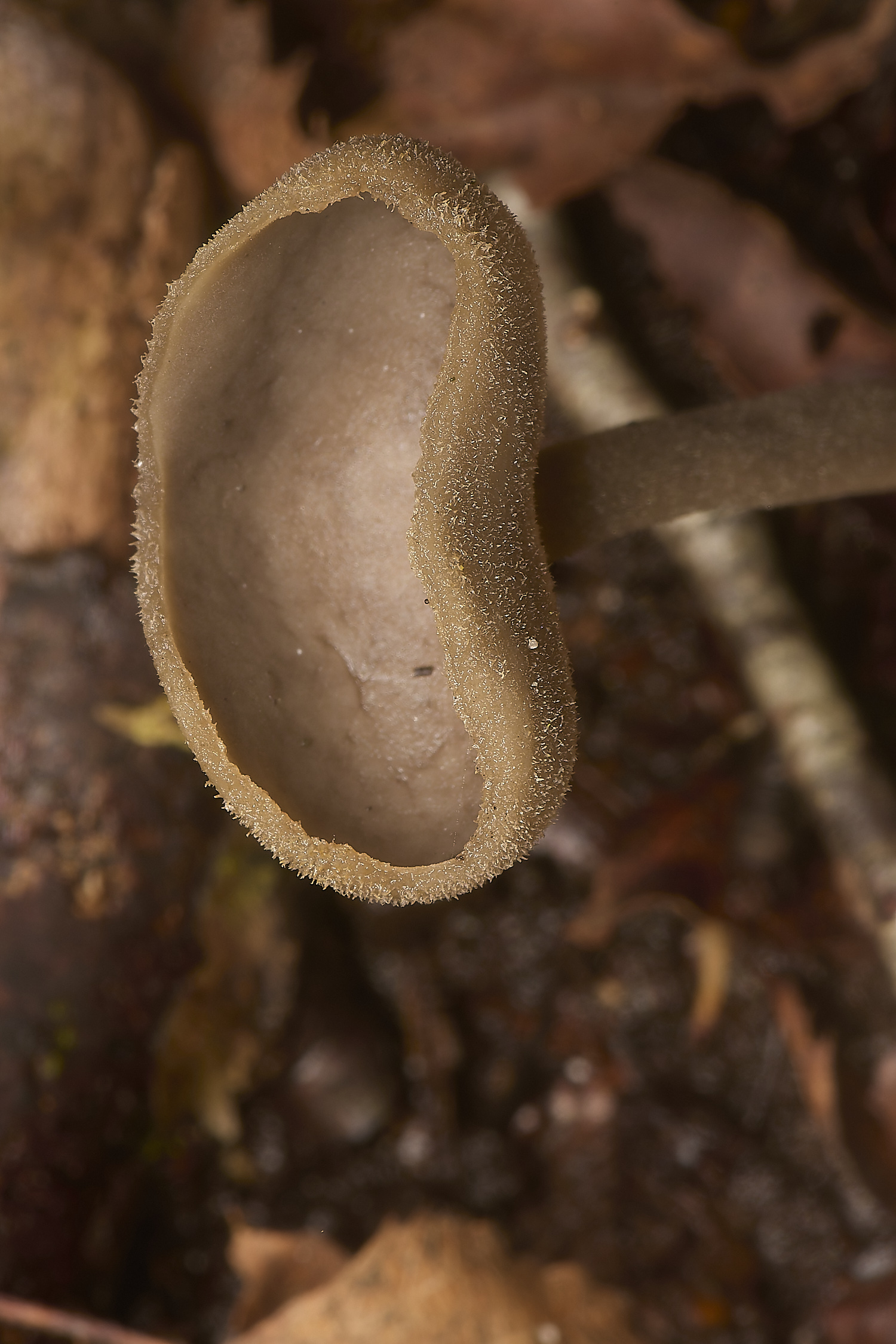
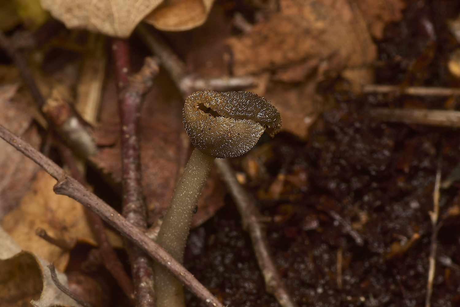
Helvella Sp
From Steve J
Starting with the Helvella, my initial thoughts when I got it home based on shape and colour was H. fibrosa AKA H. dissingii, working through Paul Cannons adaptation of the Spooner
Helvella key very quickly put it as Helvella macropus, just to be on the safe side I double checked everything using Peter Thompson, Fungi of Switzerland and Fungi of
Temperate Europe all agreed with the results of the key on every aspect so happy to go with that.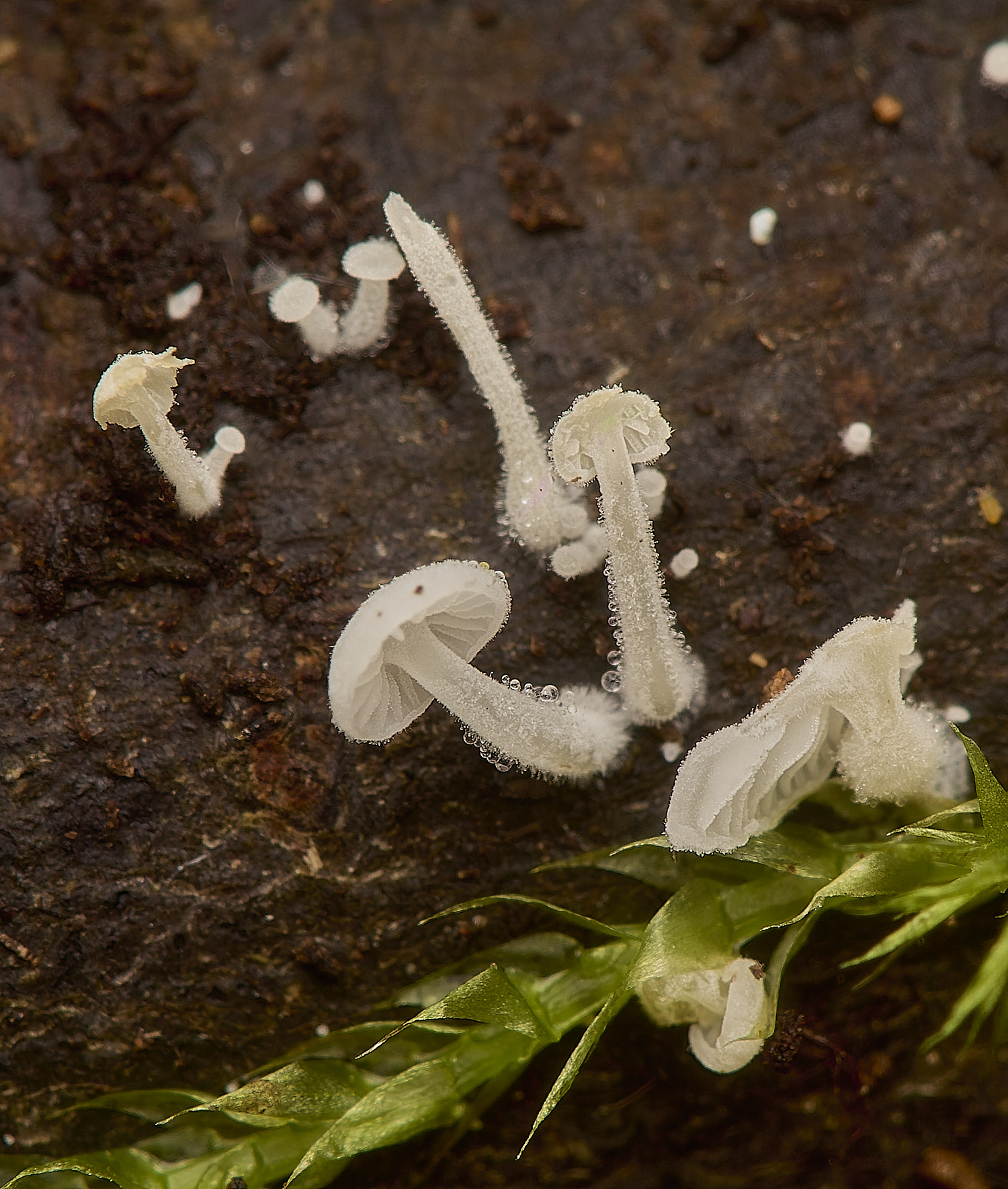
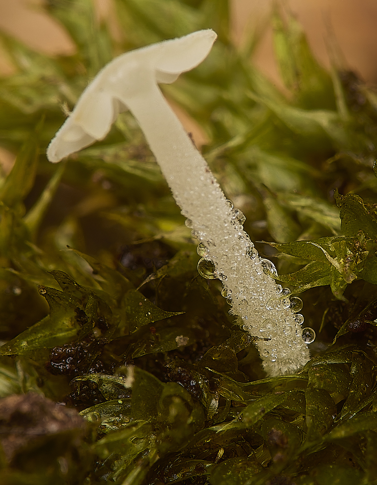
Dewdrop Bonnet (Hemimycena tortuosa)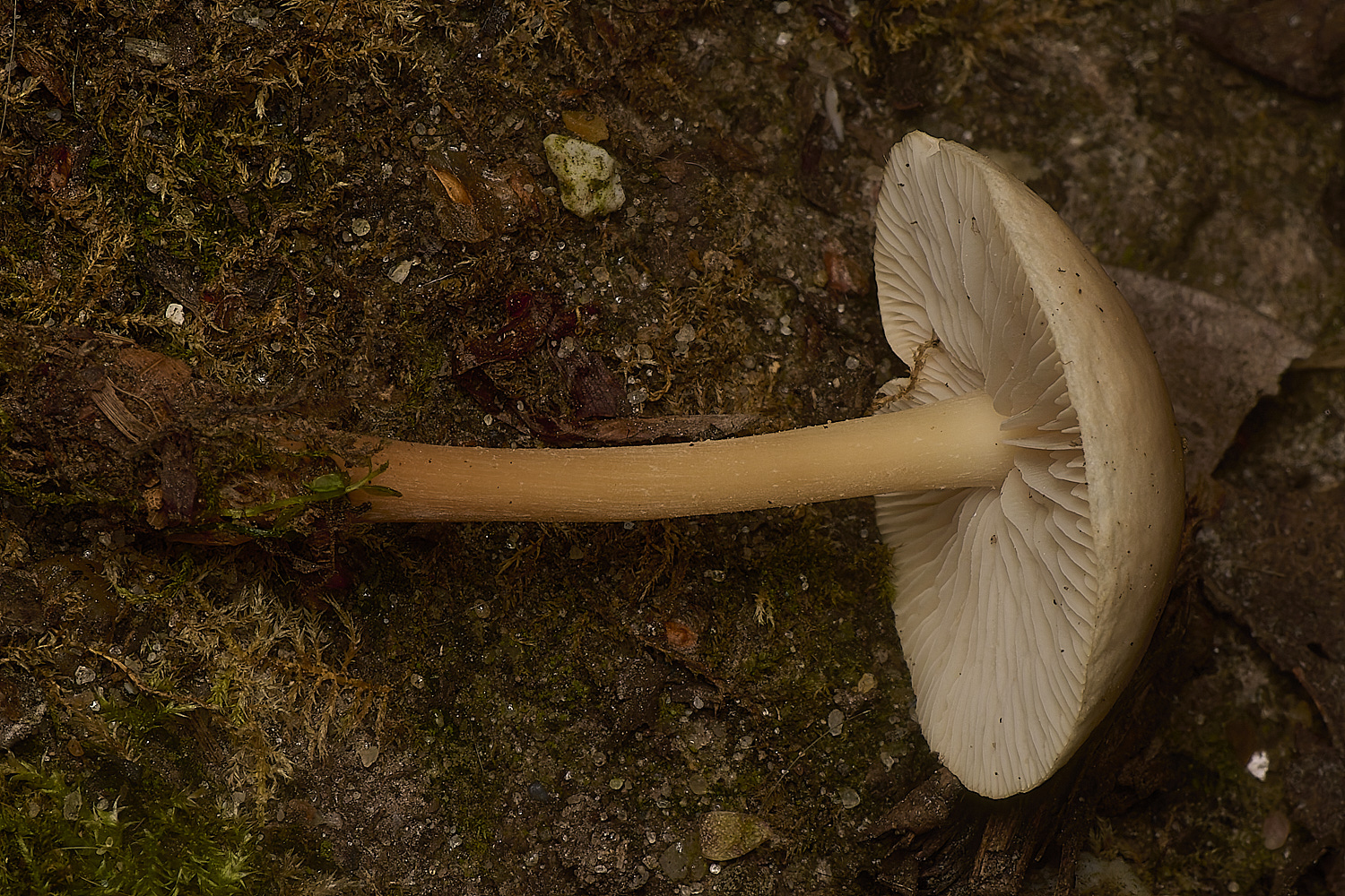
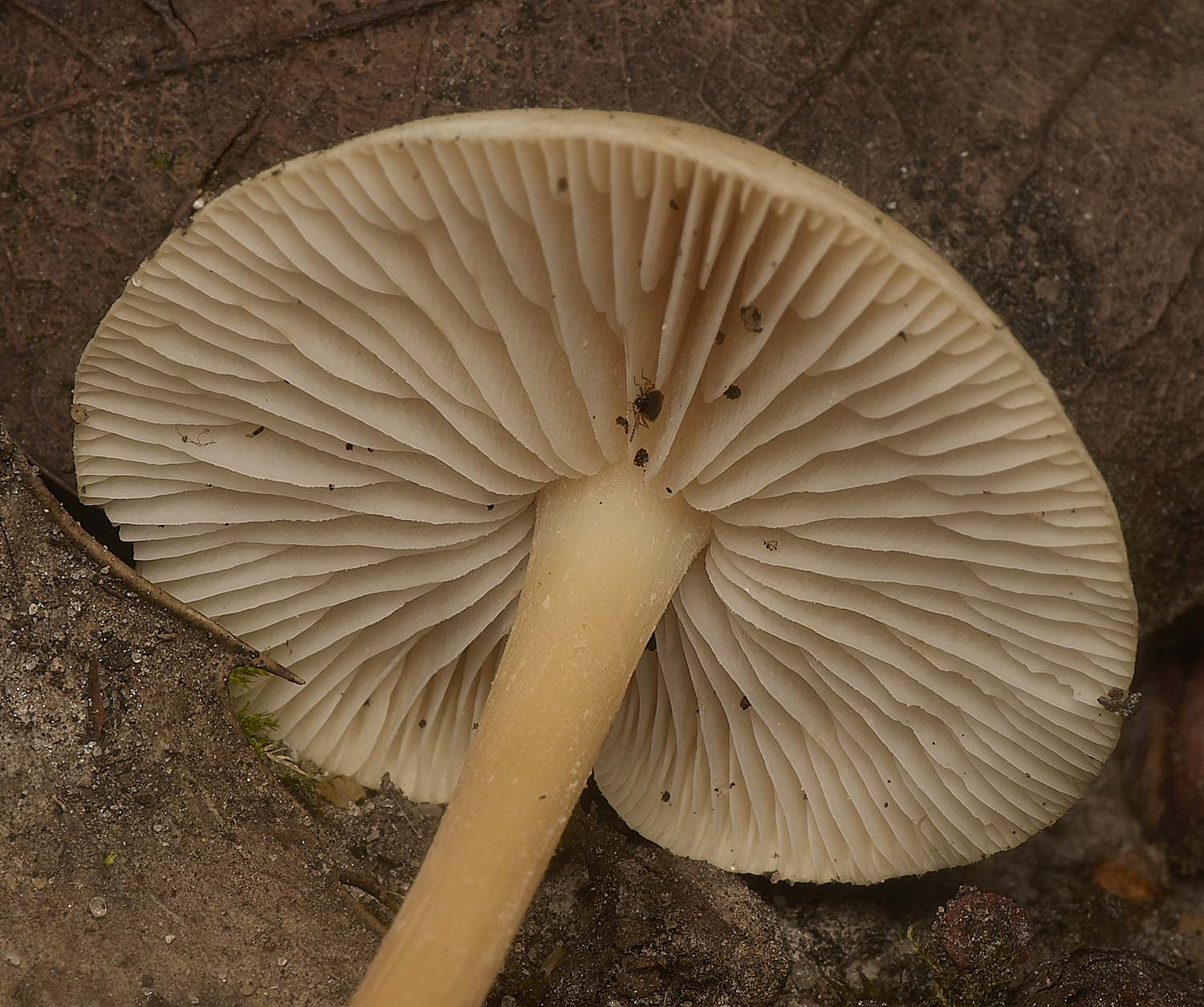
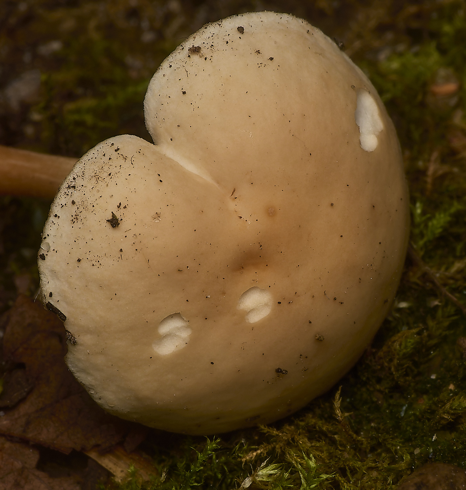
Russet Toughshank (Gymnopus dryophilus)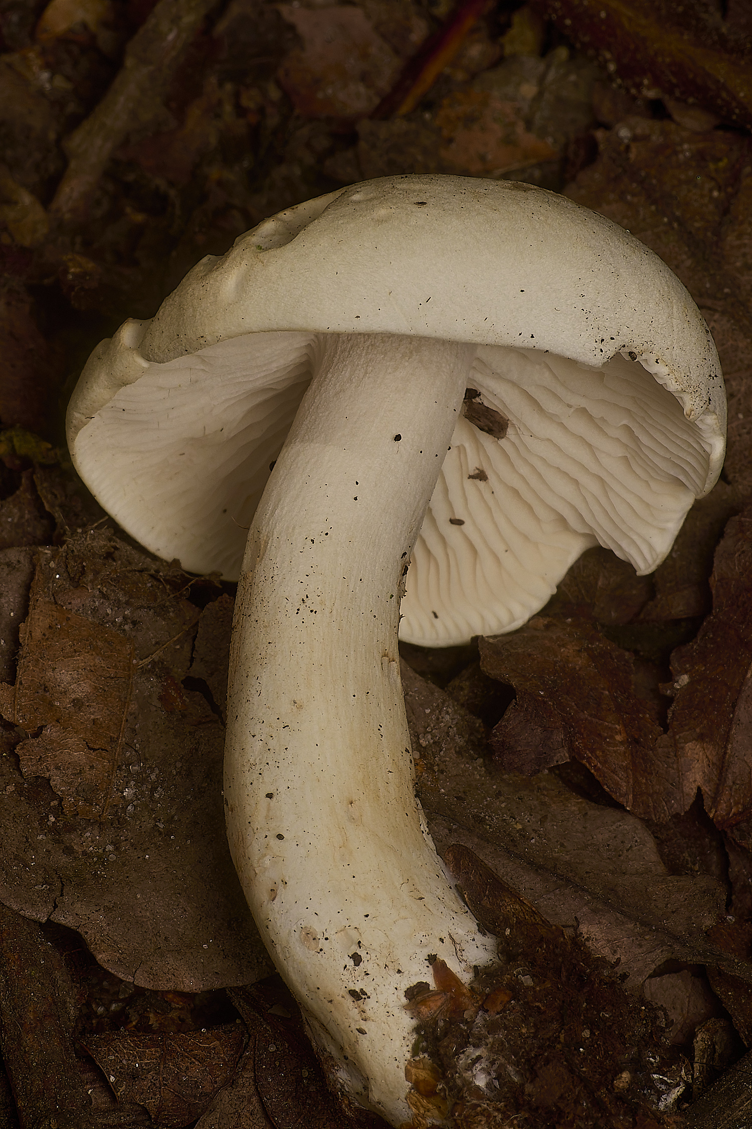
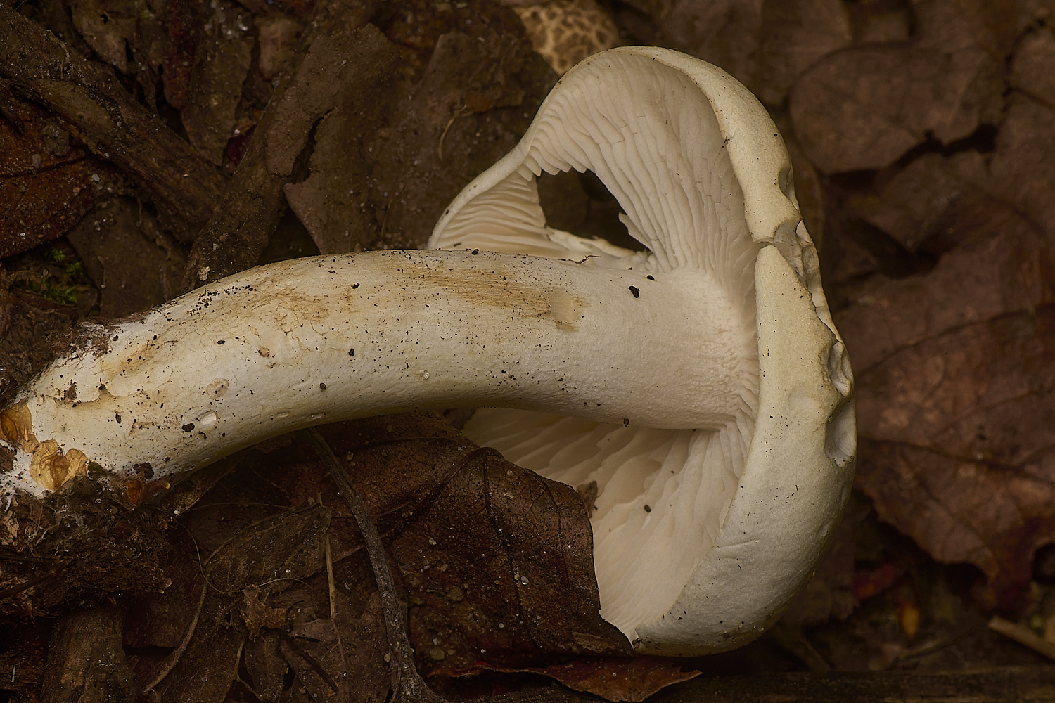
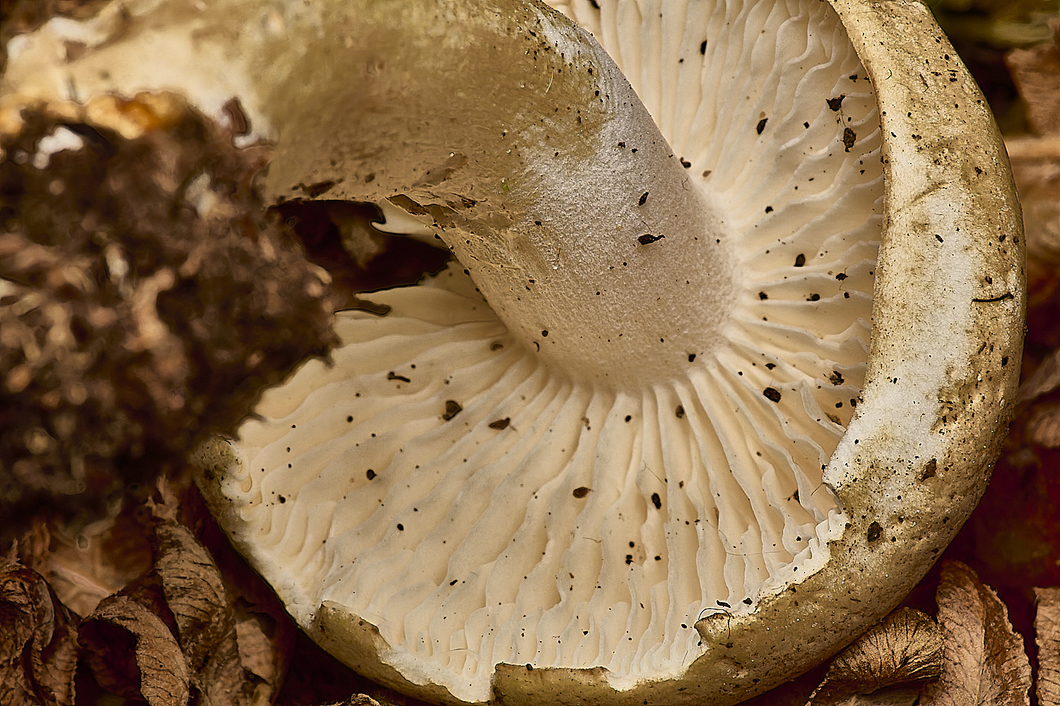
Chemical Knight (Tricholoma stiparophyllum)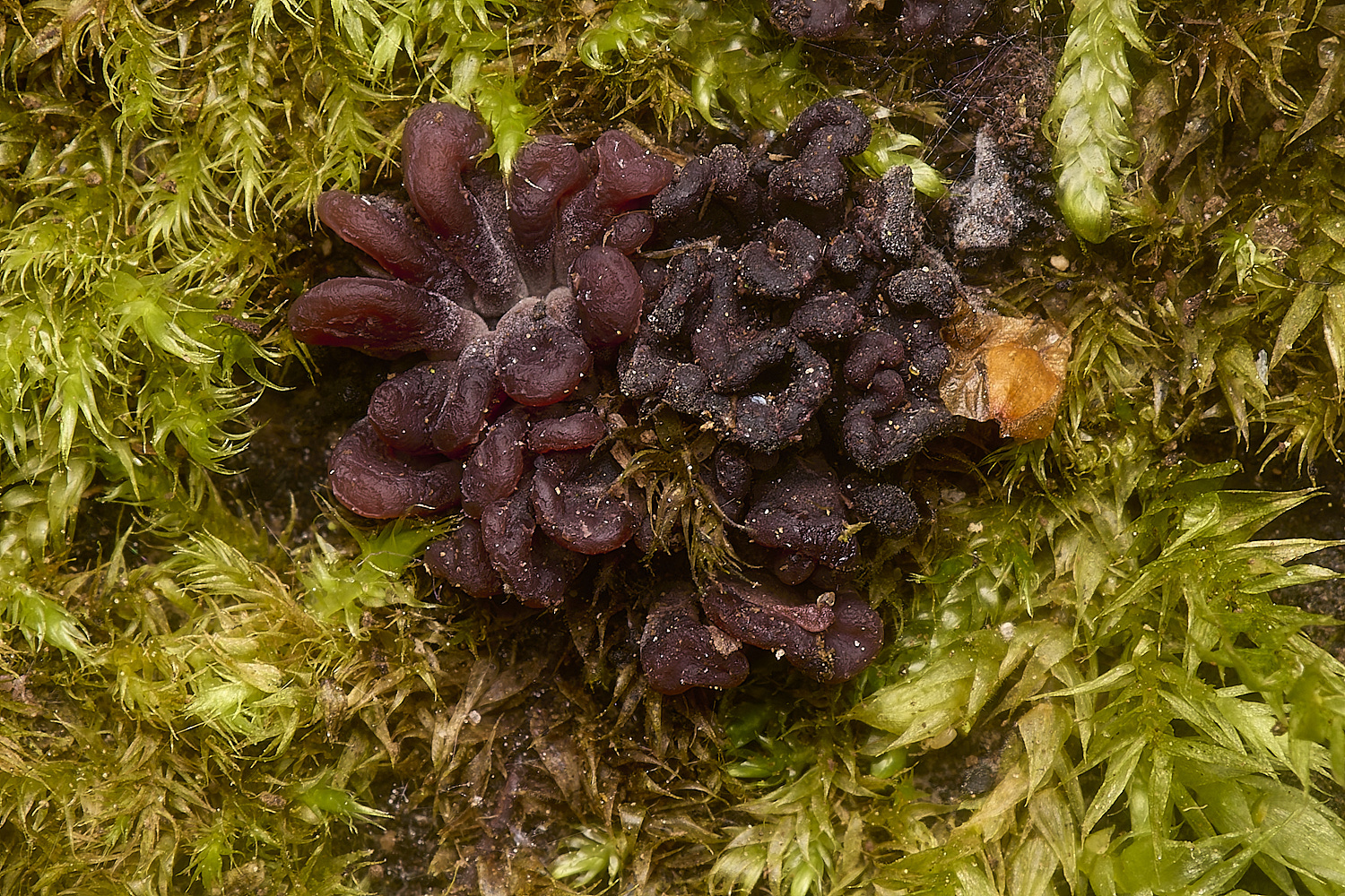
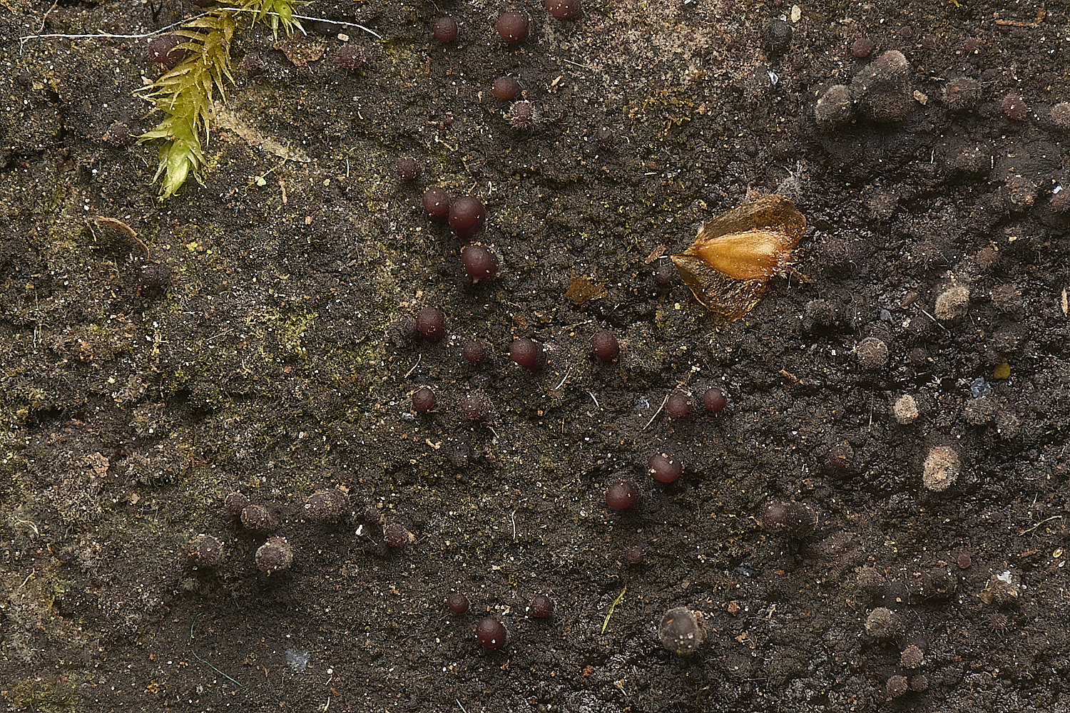
Purple Jelly Disc (Ascocroyne sarcoides)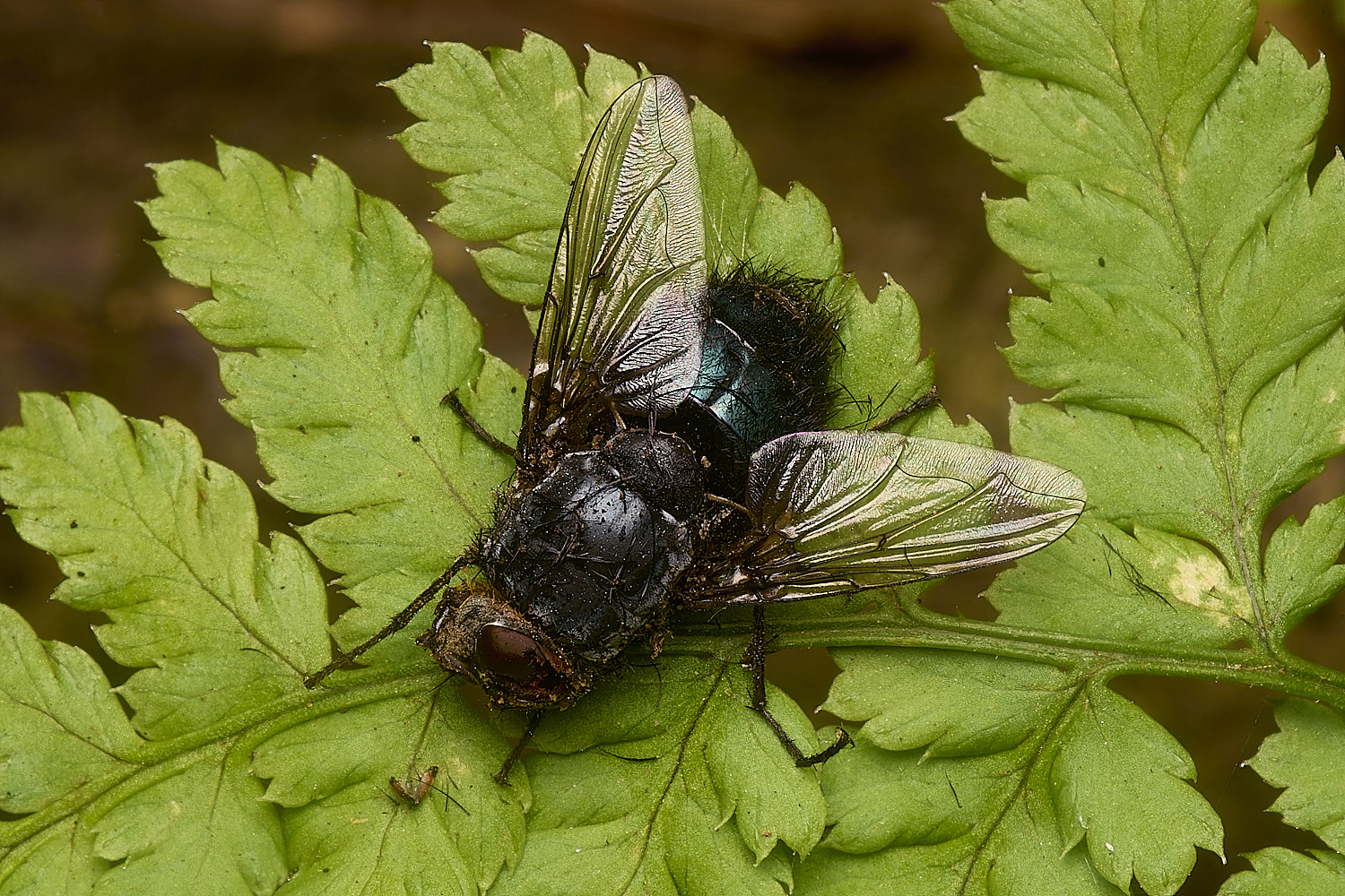
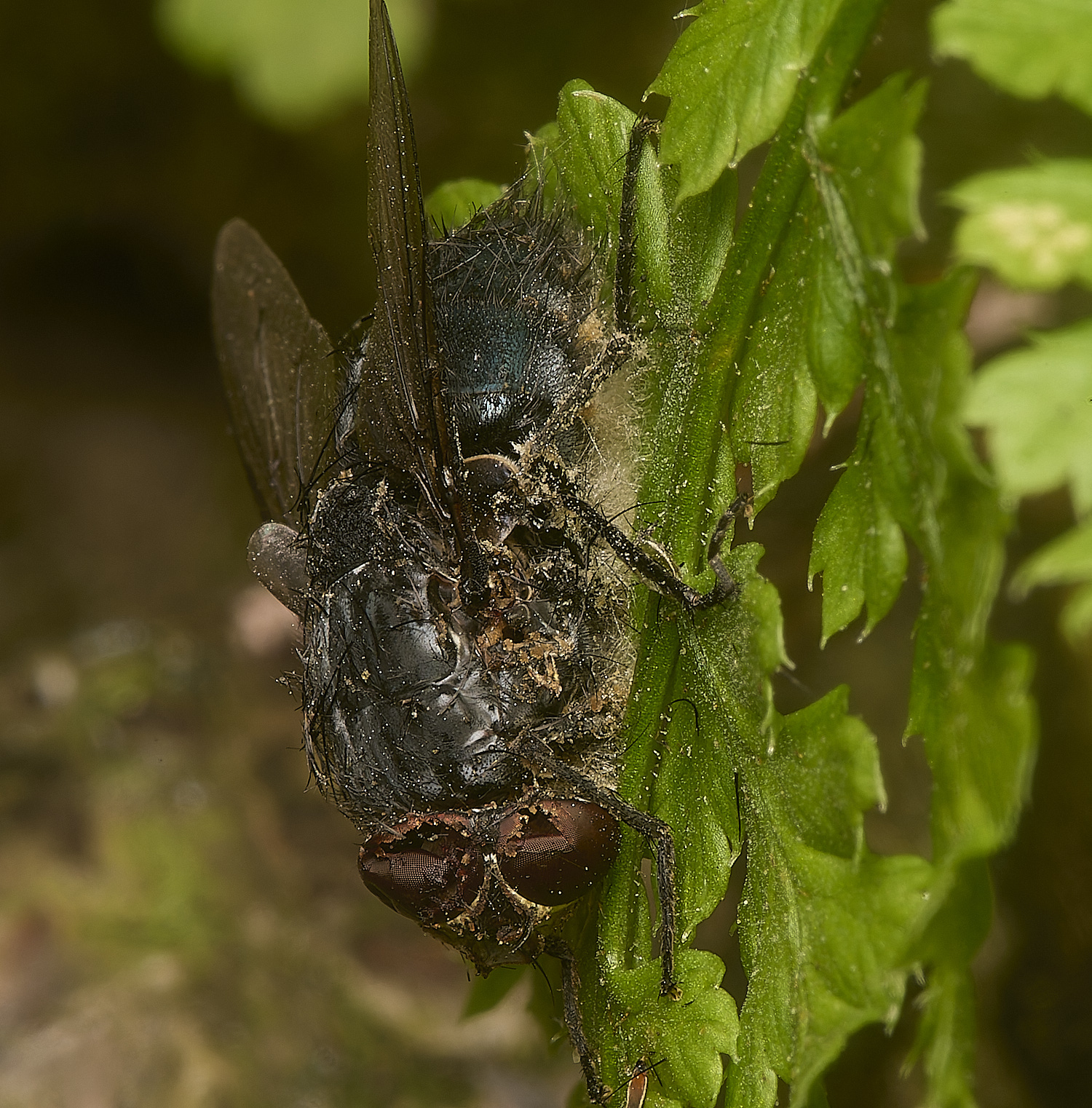
Once placed in a damp environment, the dead fly infected by an entomogenous fungi soon shed numerous conidia around the body, forming a
white mass on the slide. Using Entomopathogenic Fungal Identification, Humber. R. Nov 2005, I got to the genus Entomophthora, based on the
characteristic lemon-shaped conidia 26x17 with several (<8) small nuclei with mean diameter of c.6um contained within. Many of these primary
conidia appeared to be germinating secondary conidia. And then using Entomophthorales of Switzerland Part I, Keller. S, it keys out to
Entomophthora schizophorae. However, I note several references to the 'E. muscae species complex' which contains E. schizophorae
so any thoughts on whether we should record it as Entomophthora muscae s.l.
From Tony M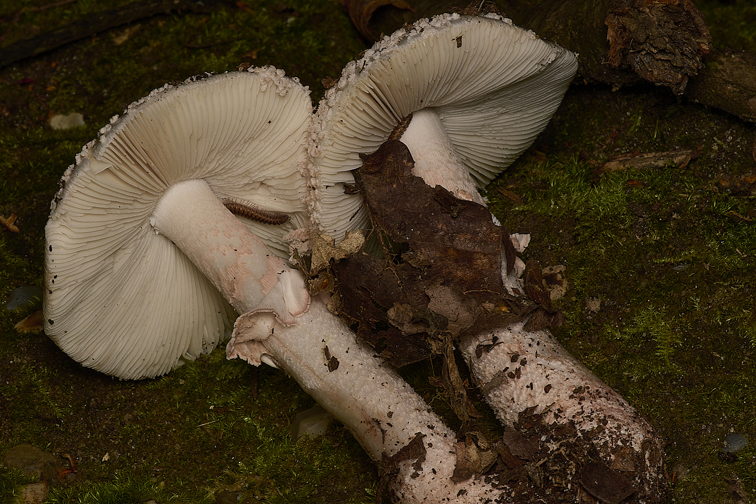
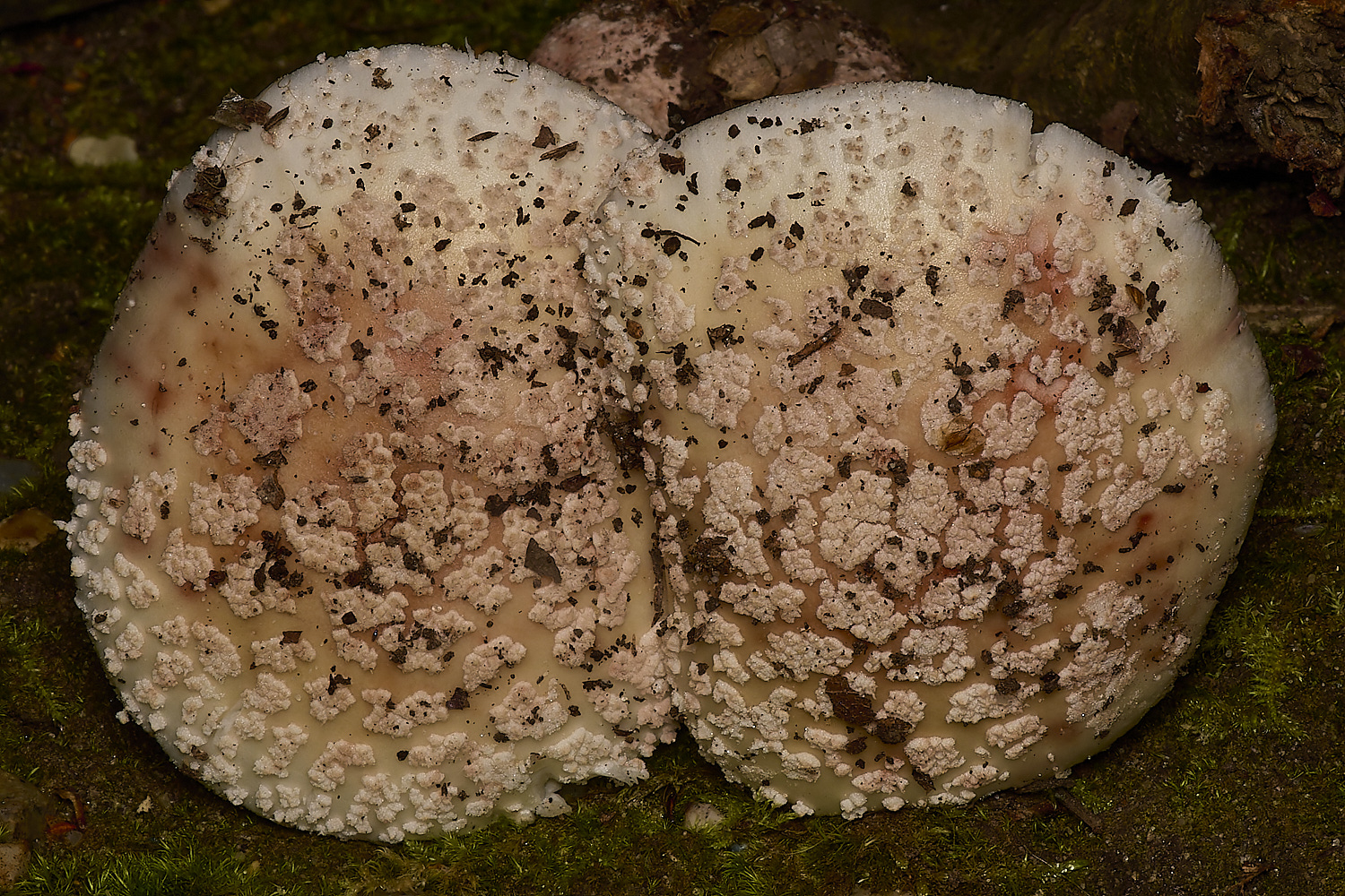
The Blusher (Amanita rubescens)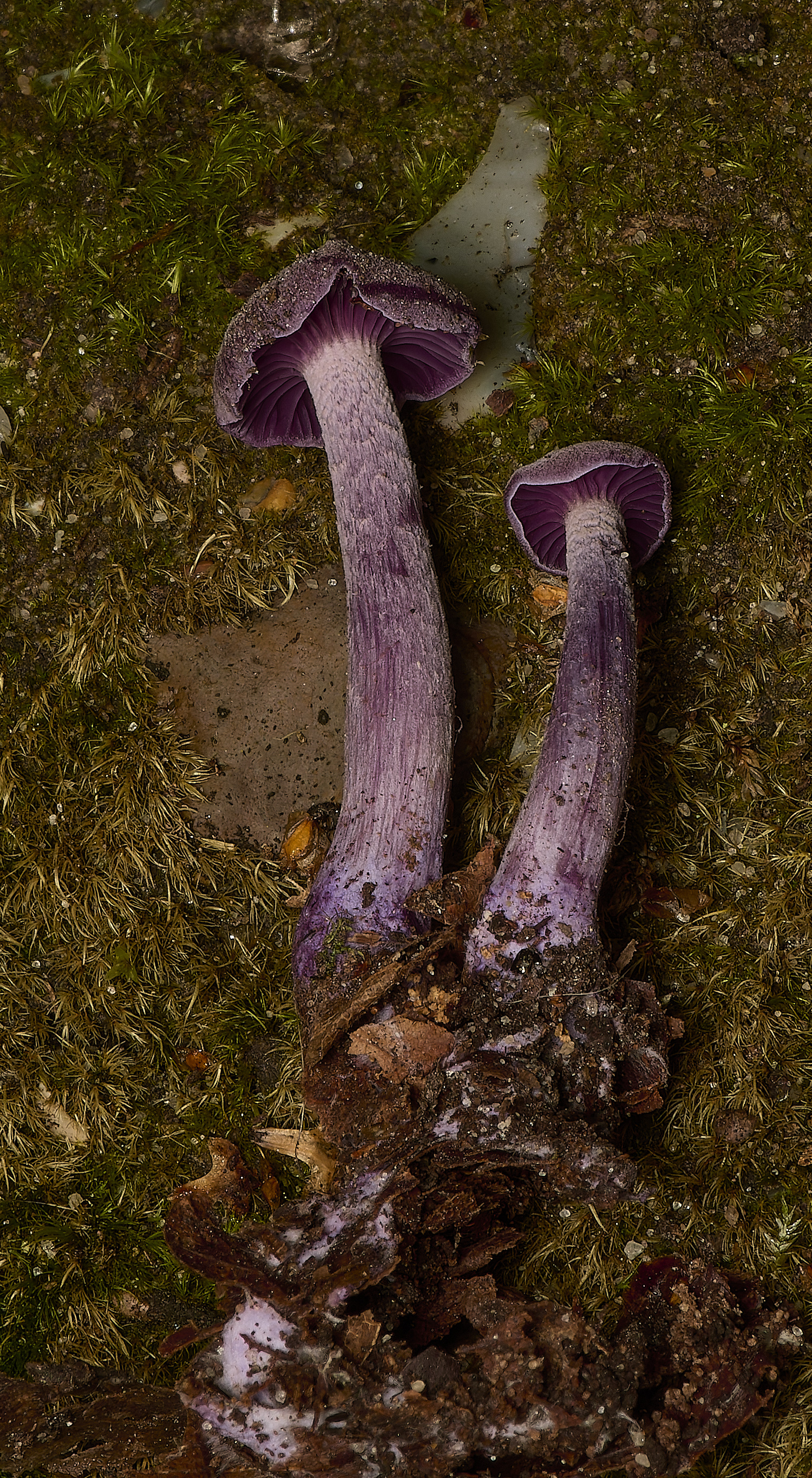
Amethyst Deceiver( Laccaria amethystina)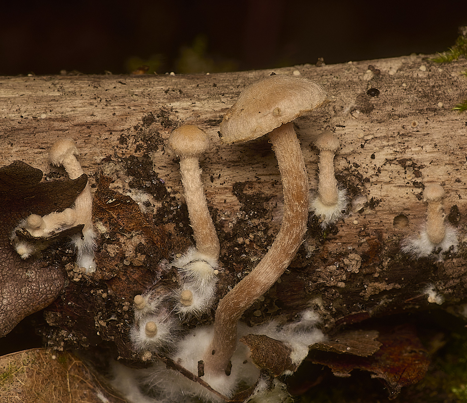
Clustered Toughshank (Gymnopus confluens)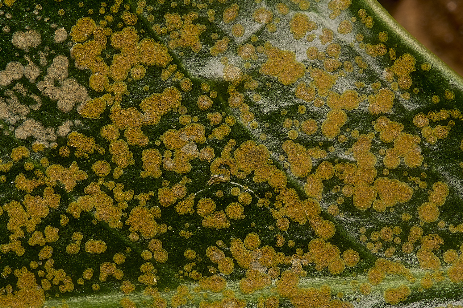
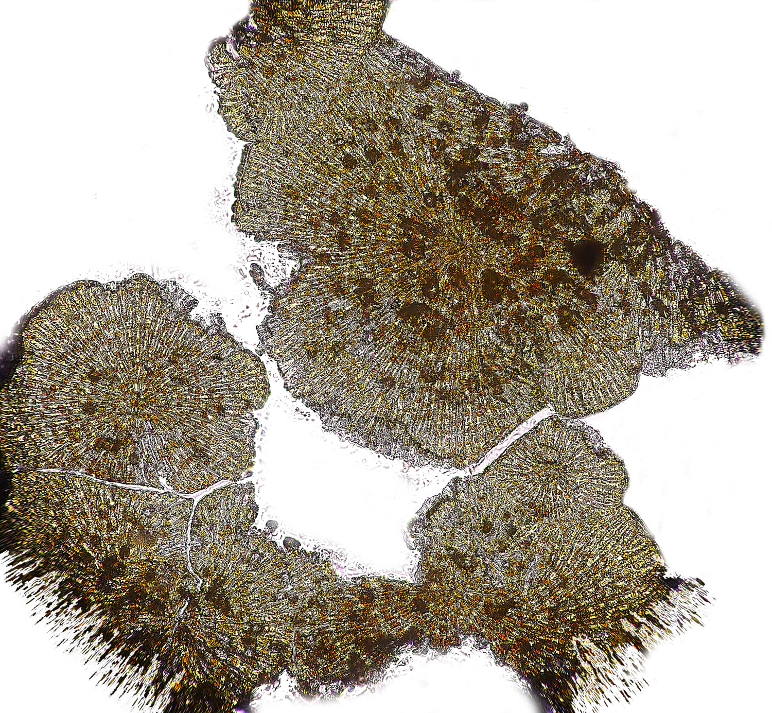
x200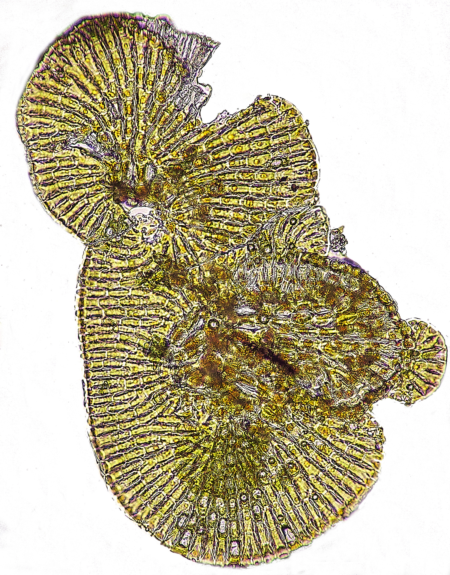
X 400
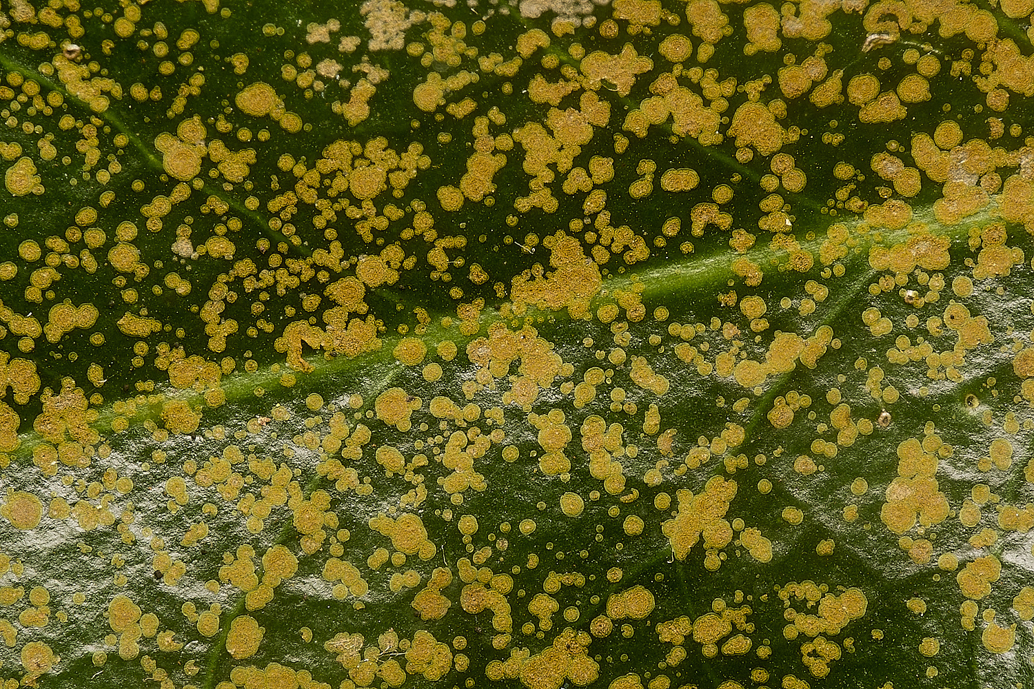
Algae Sp on Holly (Ilex aquifolium)
Phycopeltis arundinacea
Tony L sent one of the Holly leaves with the above alga on it to Faye Newberry, a plant pathologist at the RHS and received an intriguing reply
Hi Tony.
Thanks for a really exciting holly leaf. The main feature was the alga Phycopeltis arundacea.
This is common on long-lived, waxy, evergreen leaves in humid locations. It does require sunlight, so is not often found deep inside the canopy.
But did you notice the tiny white apothecia on the leaf? They were clustered around an old wound – probably a scar from a spine on a neighbouring leaf.
There was a small amount of green-grey, granular crust. This was composed of single-celled green algae with lots of fungal hyphae.
The apothecia were flat, white, 0.2mm or less in diameter, with a white margin that was difficult to see as the disc was also white.
(You’d need to make a cross-section rather than a squash.) There was no pigmentation in any of the tissues.
Asci were narrowly cylindrical, 8-spored, with walls staining with Lugol’s iodine but no tip architecture picking up the strain.
Paraphyses were hard to find and I’m not sure that I saw any! Ascospores were long and narrow, approx. 40 um x less than 2 um,
with the top end wider than the bottom half. I didn’t mount in a stain so was unable to tell with such narrow spores, whether there were any septa or not.
I sent photos and a description to Brian Coppins. He’s pretty sure this is Bacidina chloroticula. There are some photos on the FGBI website but no description.
I found a description here: https://britishlichensociety.org.uk/sites/default/files/Ramalinaceae%20rev%201a_0.pdf There are a couple of previous records for this lichen in East Norfolk the BLS database.
See https://britishlichensociety.org.uk/maps#Bacidina_chloroticula It’s a shame it wasn’t in the West as it might have been a new county record – the BLS has no West Norfolk records! With your permission
I’ll include the record in my next submissions to the BLS database.
So… would you like the leaf back? There are three apothecia remaining!
I really enjoyed this verification! Thank you.
All the best, Fay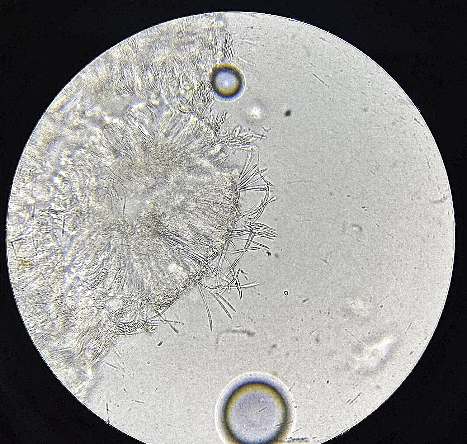
Lichen Sp
Bacidina chloroticula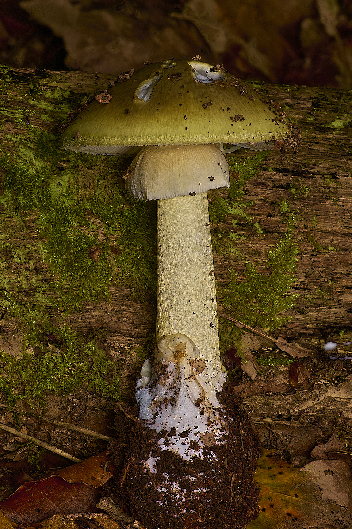
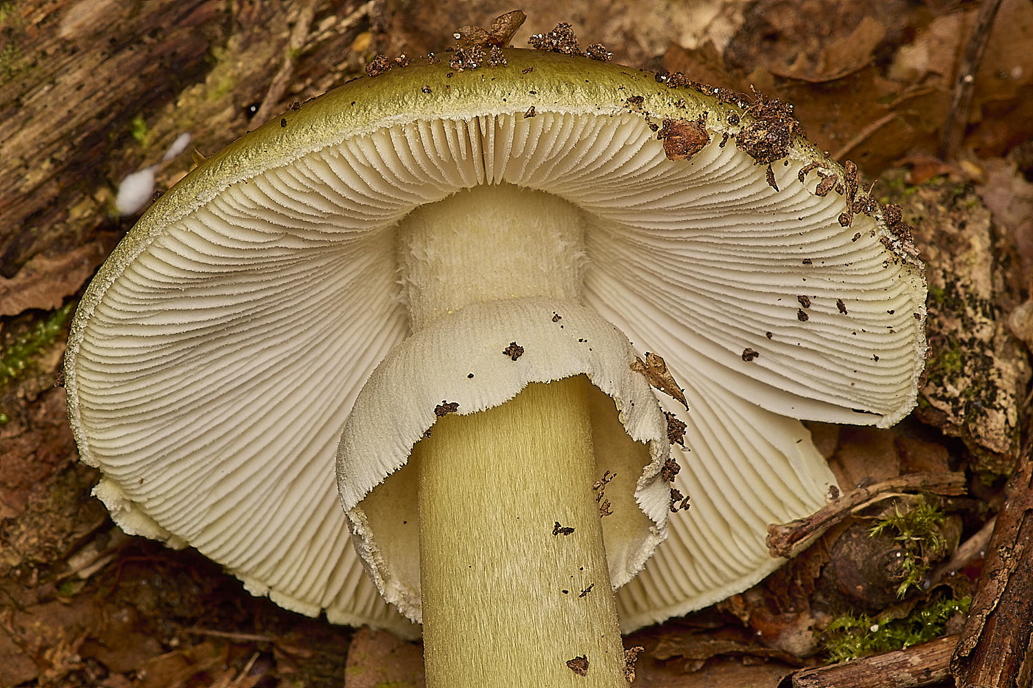
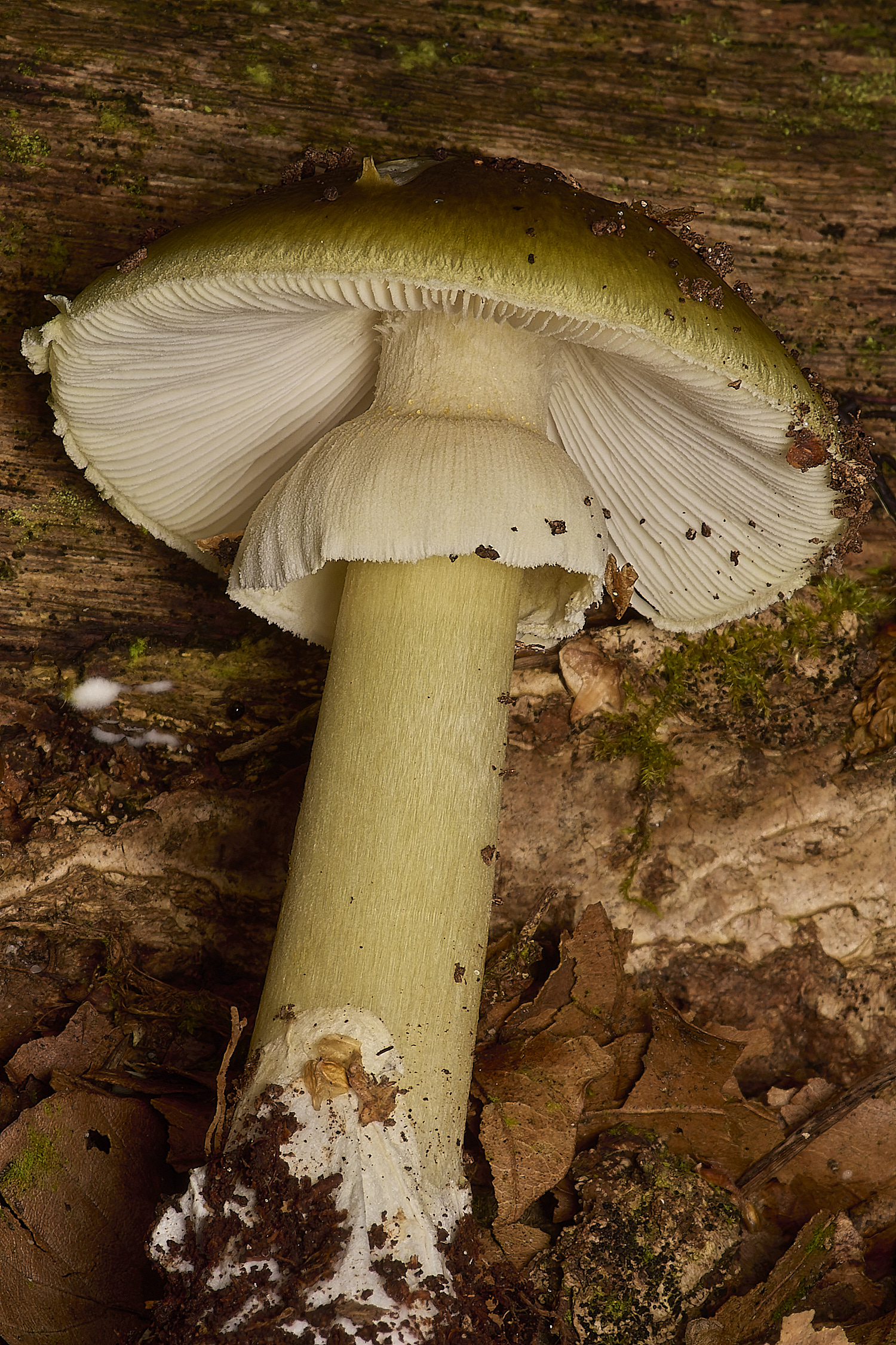
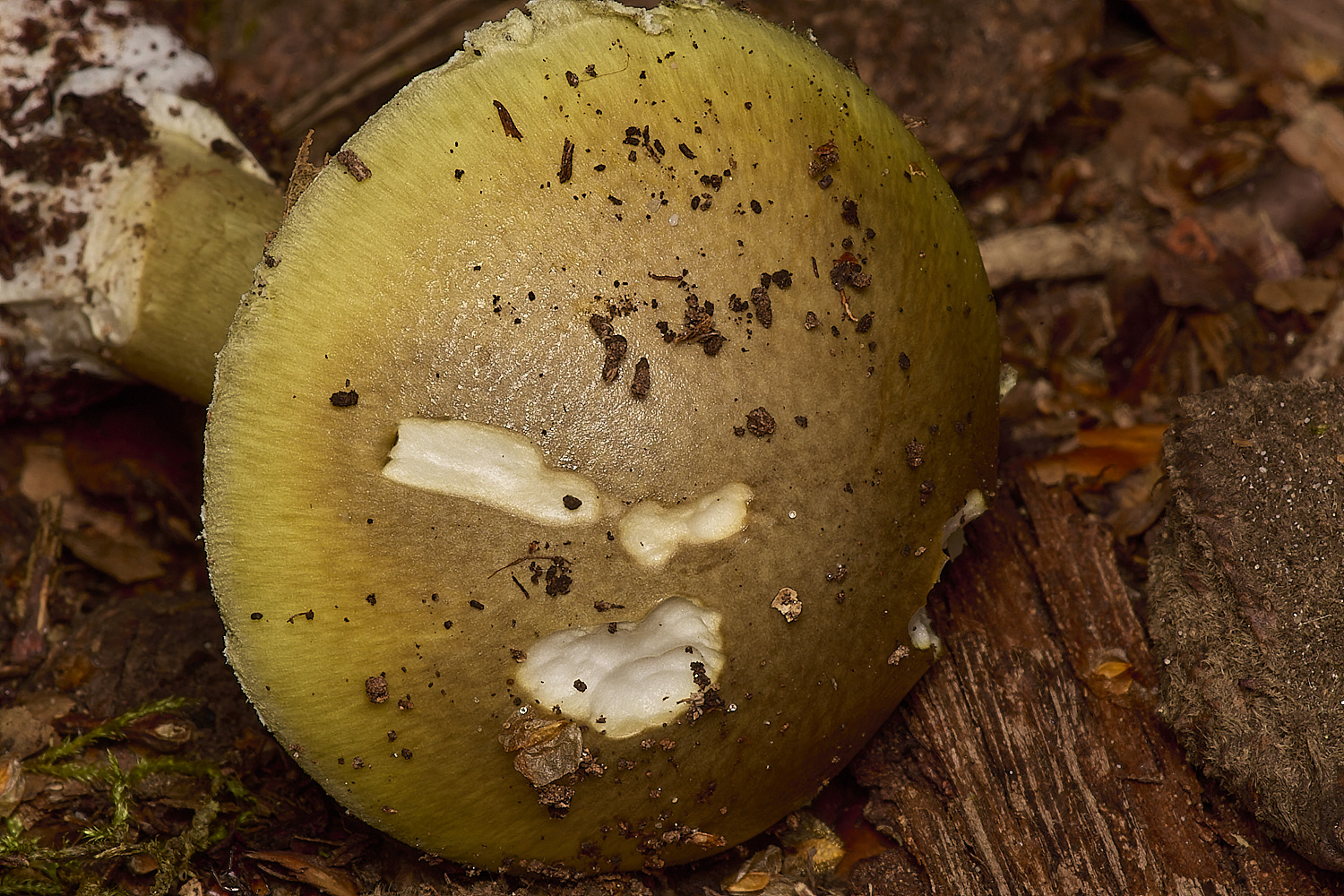
Death Cap (Amanita phalloides)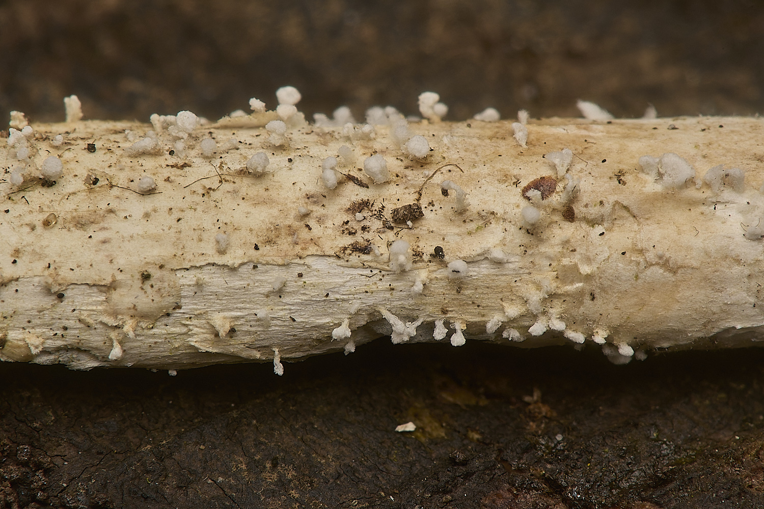
?
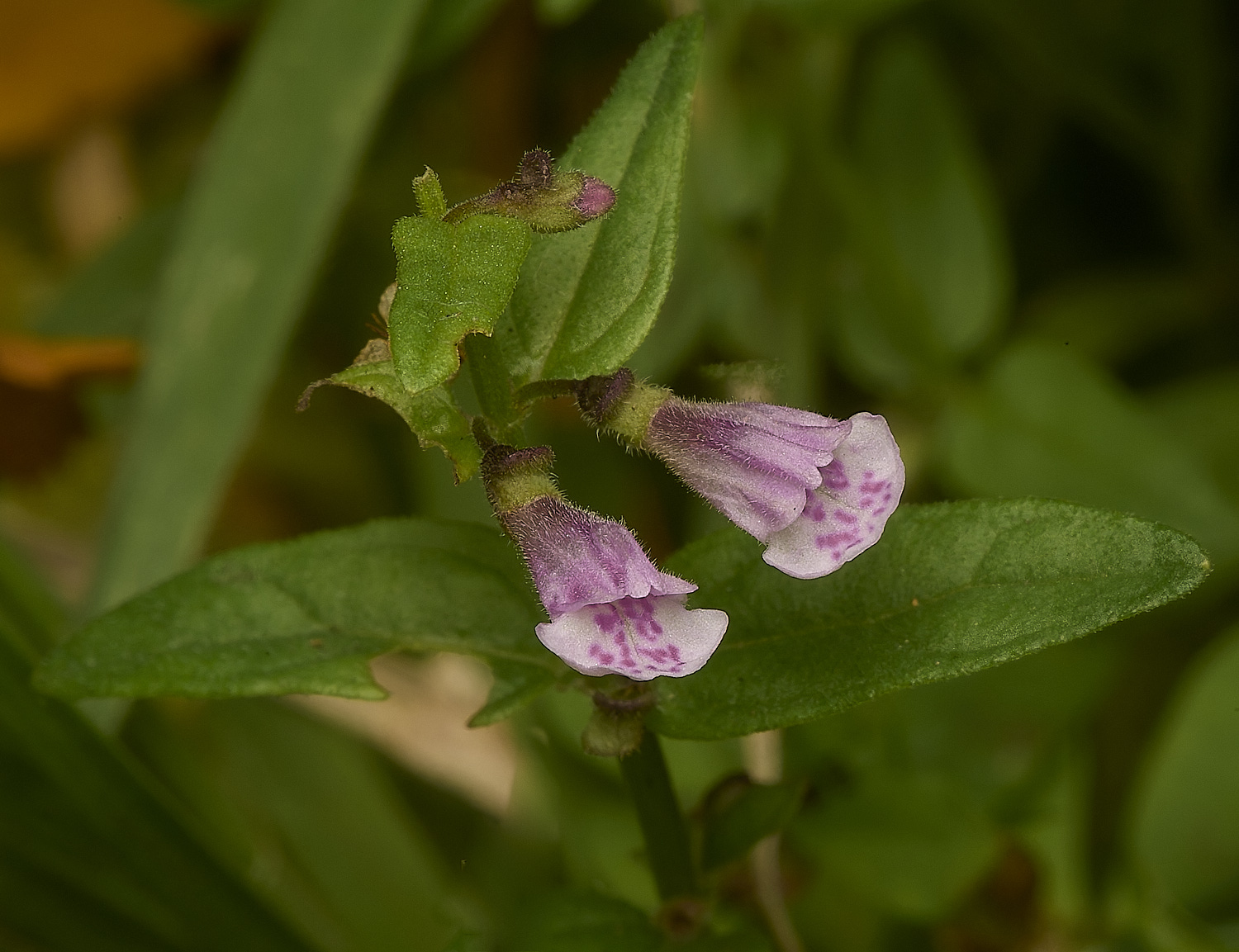
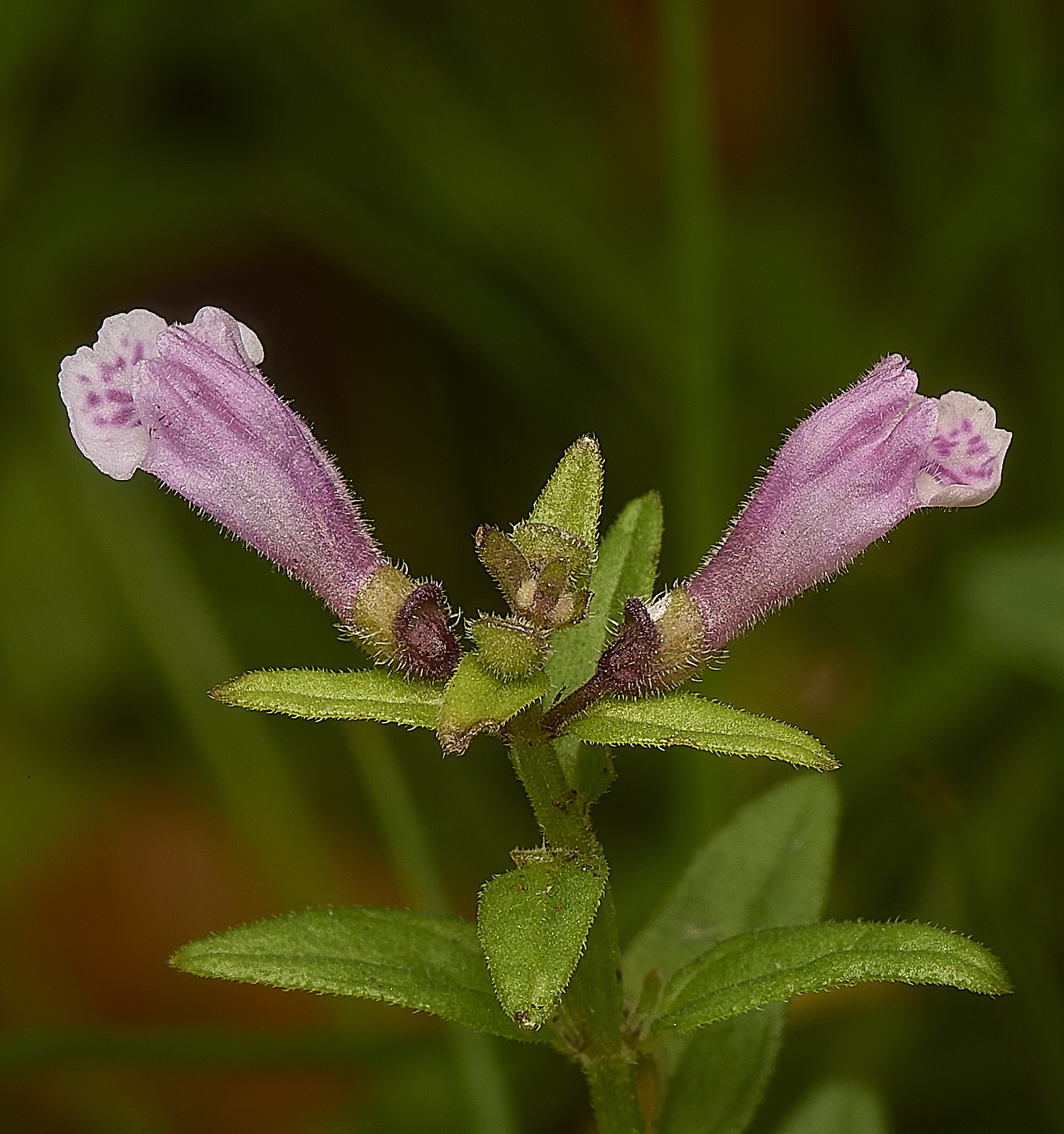
Lesser Skullcap (Scutellaria minor)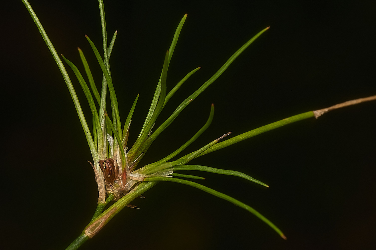
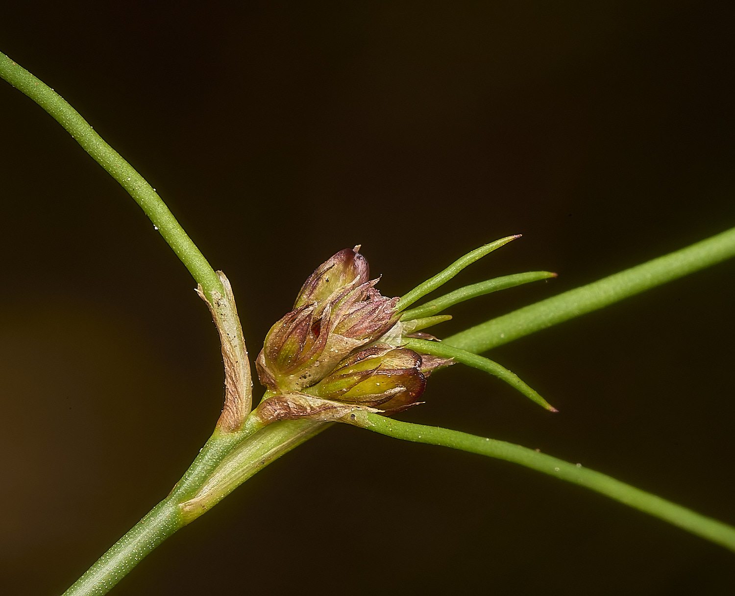
Bulbous Rush (Juncus bulbosus) exhibiting its viviparous tendencies
Trilobed fruits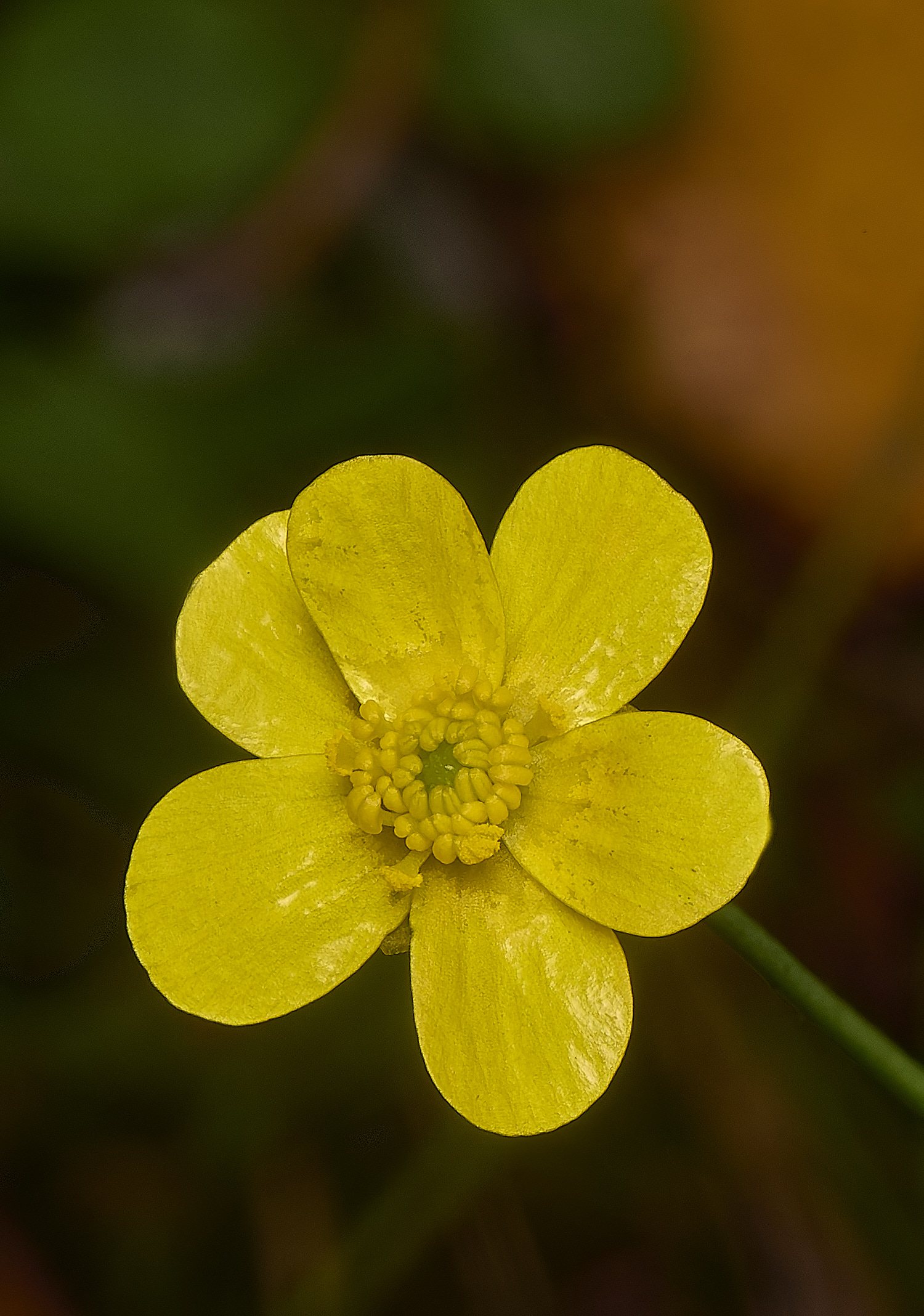
Lesser Spearwort (Ranunculus flammula)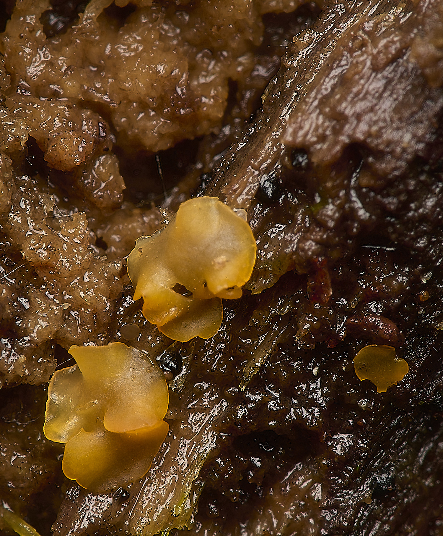
Orbilia Sp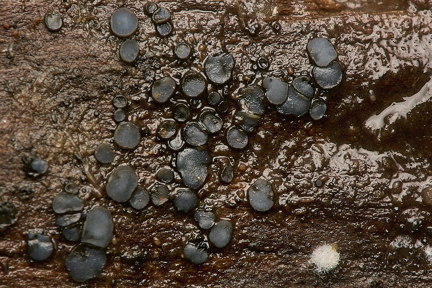
Mollisia Sp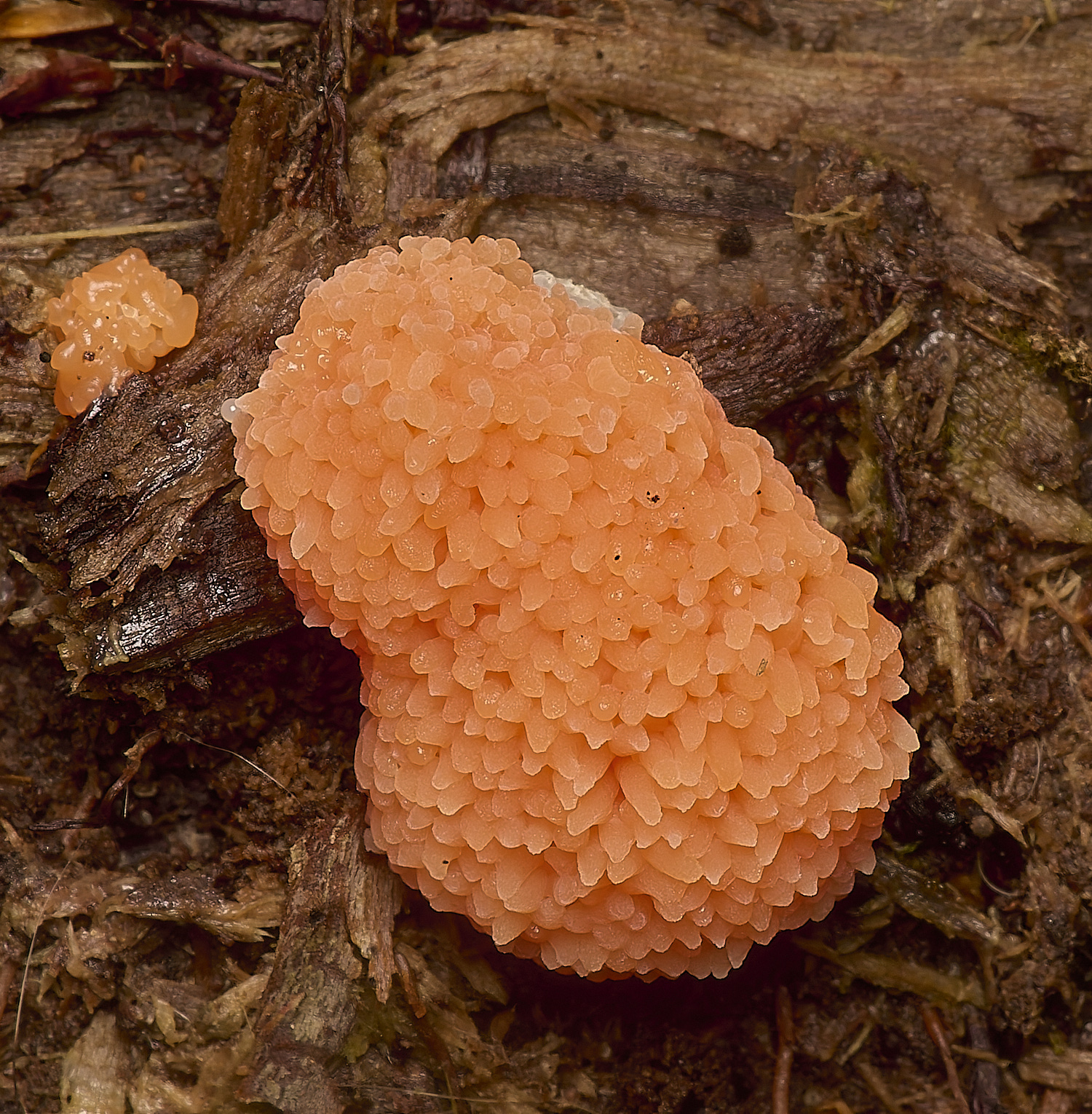
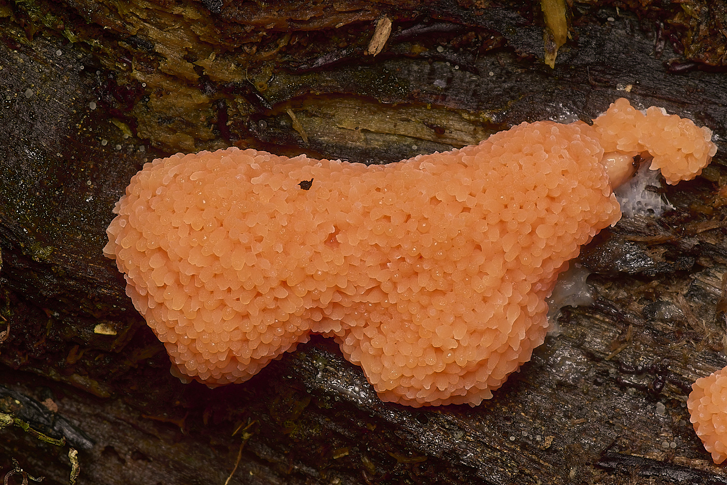
Raspberry Slime Mold (Tubifera ferruginosa)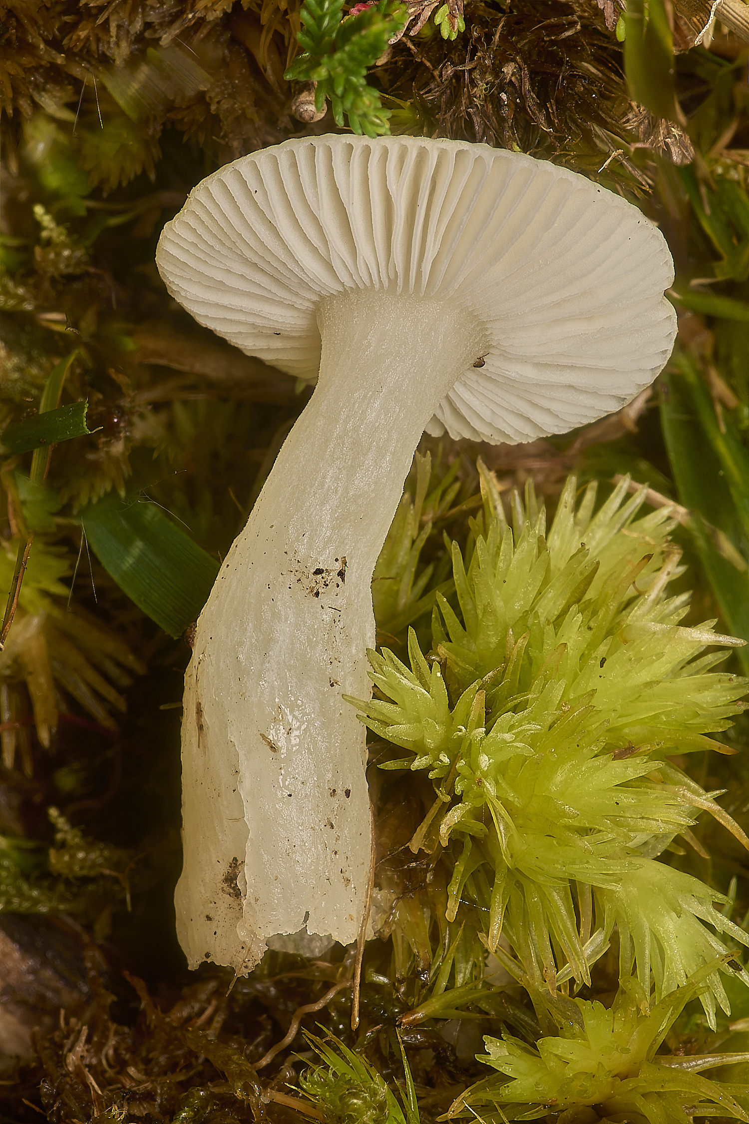
Snowy Waxcap (Cuphophylus virgineus) with an accompanying tuft of Pincushion Moss (Leucobryum glaucum)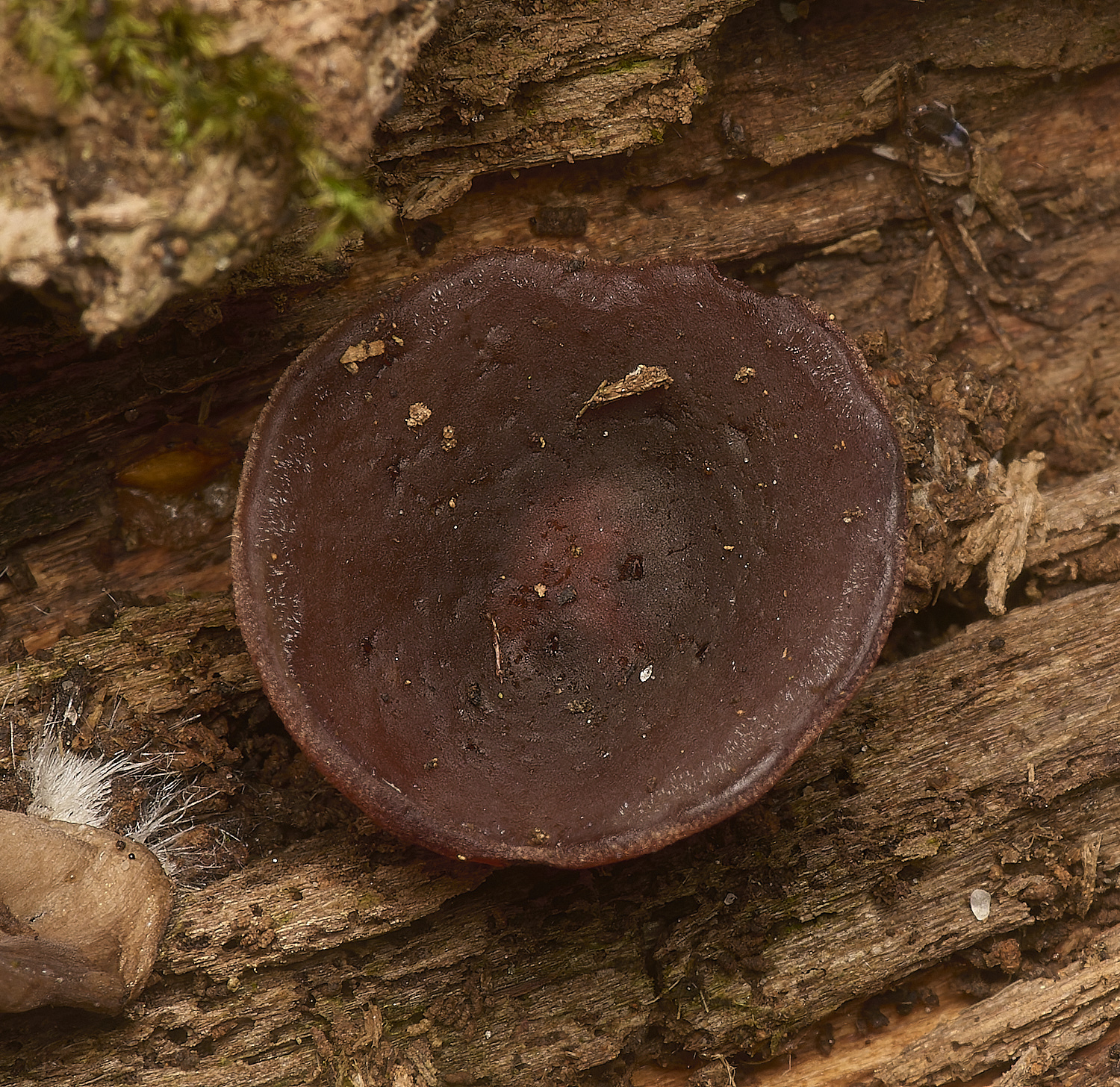
Brown Cup Fungus (Peziza phyllogena)?
From Steve J
Brown Cup was neither quick nor easy. I have spent large chunks of the past couple of days on this one.
Everything I am looking at so far is causing me a great deal of uncertainty as to whether it is or not although based on spores I am inclined towards not,
I appreciate that I could well be completely wrong and am likely to have missed something obvious or have never seen Brown cup spores at this stage.
The fruit body on unidentified decayed wood was deep brown, with a hint of purple, cupulate and very short to not stemmed, 15mm x 6mm
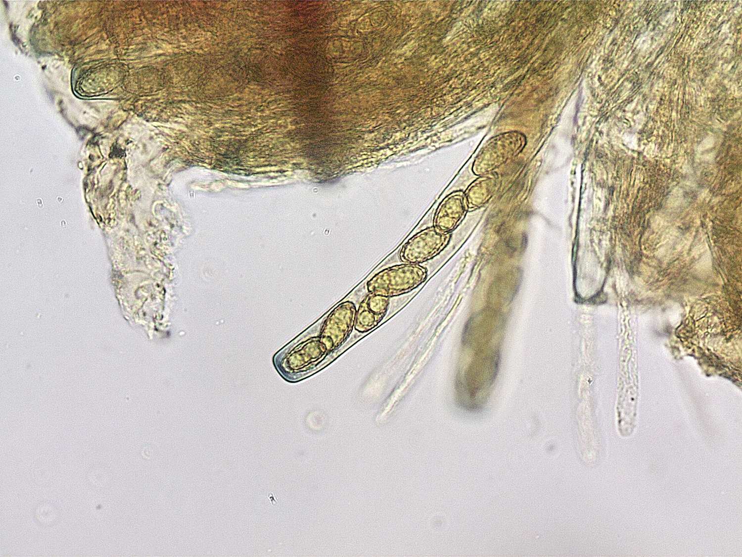
Microscopically, the spores were elliptical, multi guttulate, non septate and appeared to my eyes to be verrucose. They were 16-18 µm x 7-8 µm, with
the great majority being 16 x 7 µm. At the extremes, one was 13 x 6 µm which I shall ignore and three were 18 x 9 µm.
Ascus 8 spored, unicerate and 198 µm long, slightly amyloid at the tips, although saying this I suspect that my Meltzers is due for replacing.
Paraphyses, filiform unforked and with a slight clavate swelling at the tips.
I have worked through Ellis & Ellis, Peter Thompson, Fungi of Switzerland and Fungi of Temperate Europe and am still no further forward.
I have attached a couple of photos and would be immensely grateful for any thoughts or suggestions.
From Tony M
My only contribution to your Brown Cup mystery would be to see whether you can get some mature spores to drop naturally,
inverting the cup in a damp environment and see whether those spores look any different to those developing within the asci.
From Yvonne M
I missed this on the foray so have you got a photo of the actual fungus ? The spores do look verrucose but I think
Tony is right in that better to see them mature when out of the ascus.It’s not a Peziza type thing is it ?
They can have ornamented spores.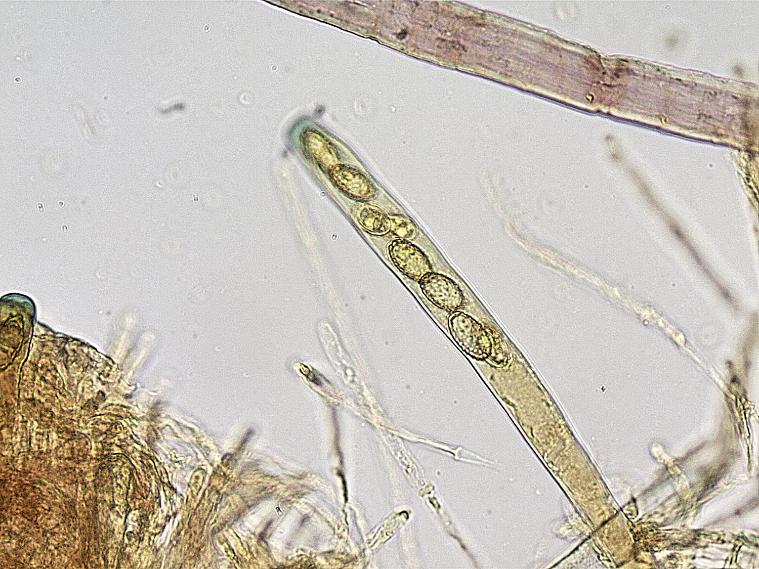
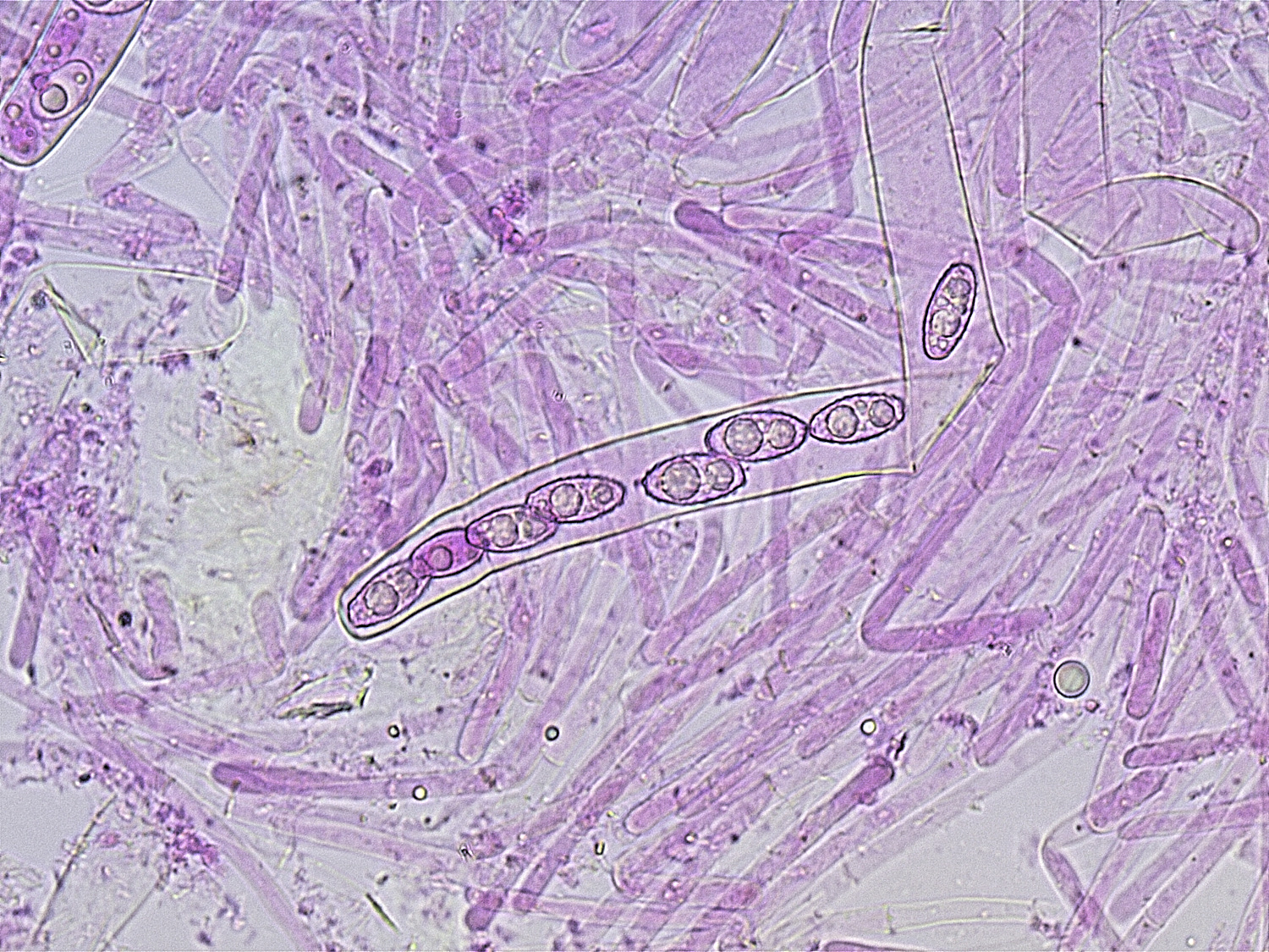
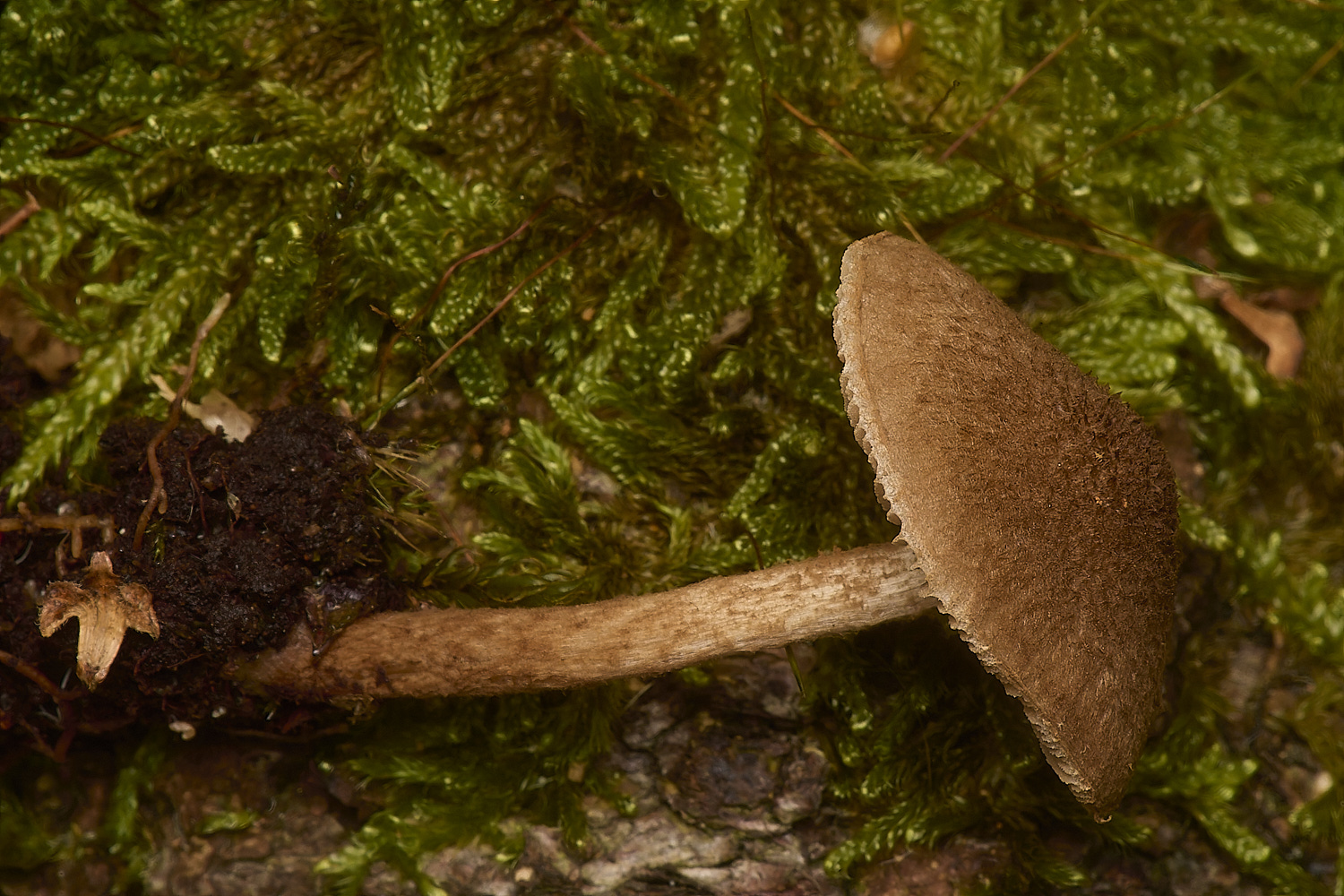
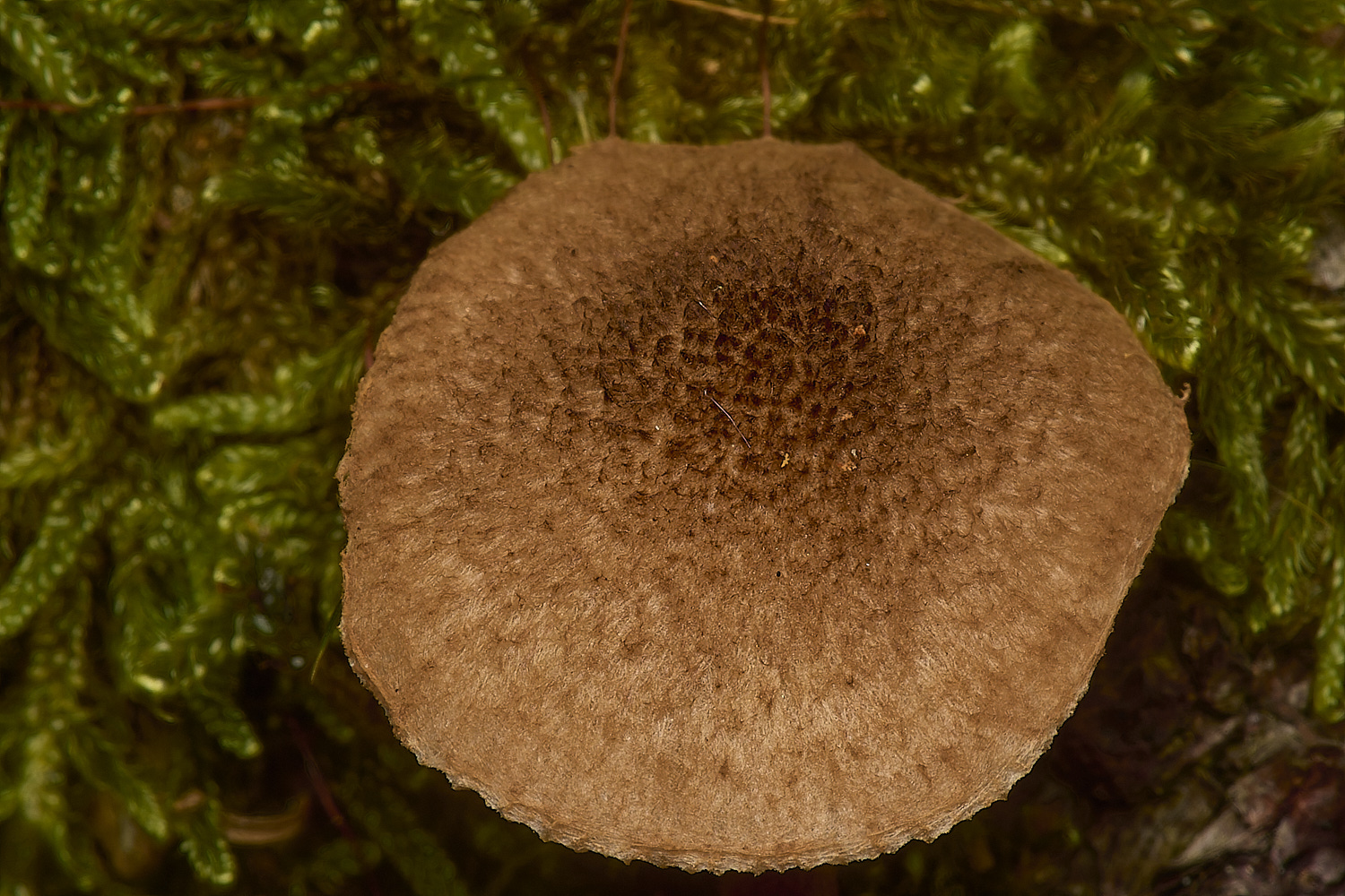
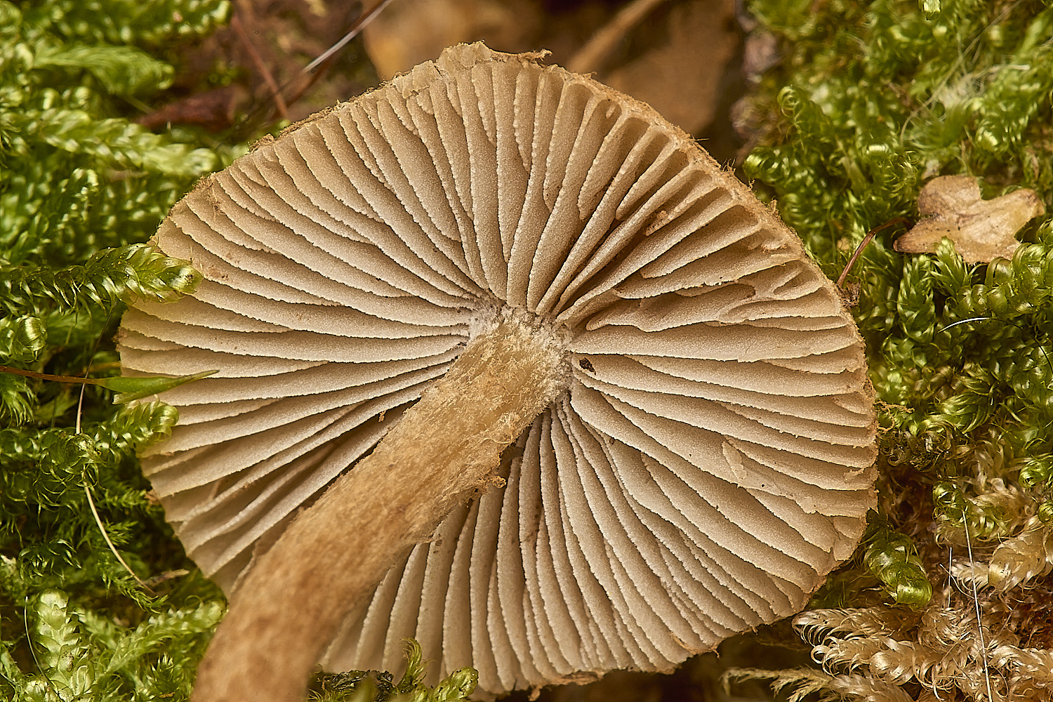
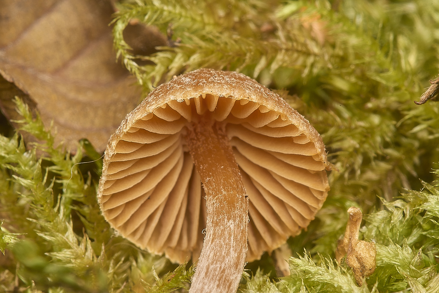
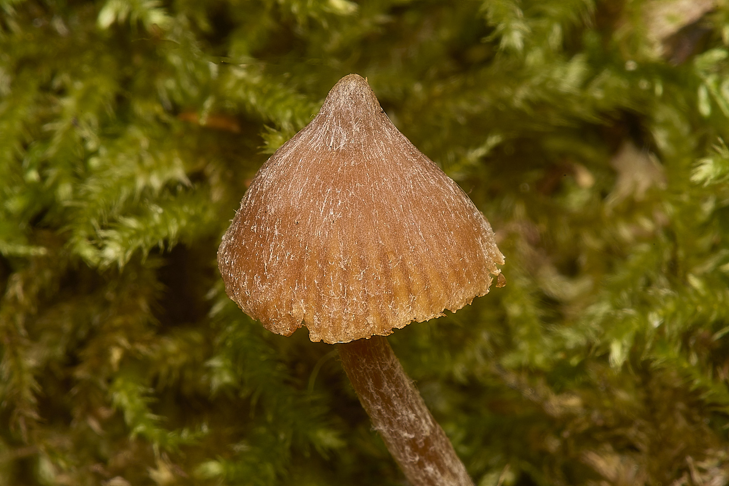
Inocybe Sp
From Tony
The brown fibrecap found towards the SE end of the wet woodland area: Using Kibby Vol 4, very knobbly (but not star-like spines) spores 10x7.2 gets you to c. 3
taxa distinguishable by shape of gill-edge cheilocystidia and presence/absence of gill-face pleurocystidia. I couldn't find any pleurocystidia so came to Inocybe leptophylla
but obviously would prefer an identification that isn't based on absence of a feature. Inocybe leptophylla hasn't been recorded in Norfolk since 2001 so its over to Yvonne to determine what this really is!
A reply from, Yvonne M
The other I hadn’t looked at when Tony M came up with an answer so probably saved me ages. I just needed to looked for pleurocystidia and I couldn’t find them either.
Like Tony I don’t like identifying anything with a negative character but in the past I have seen pleurocystidia fairly easily in the look-a-like I. stellatospora.
As this seems to be the only way to separate the 2 I think we should go with I. leptophylla. Also, the cheilcystidia do look more like this than those of stellatospora
as they seem thin walled. Penny Cullington’s key, Stangl and Kibby vol 4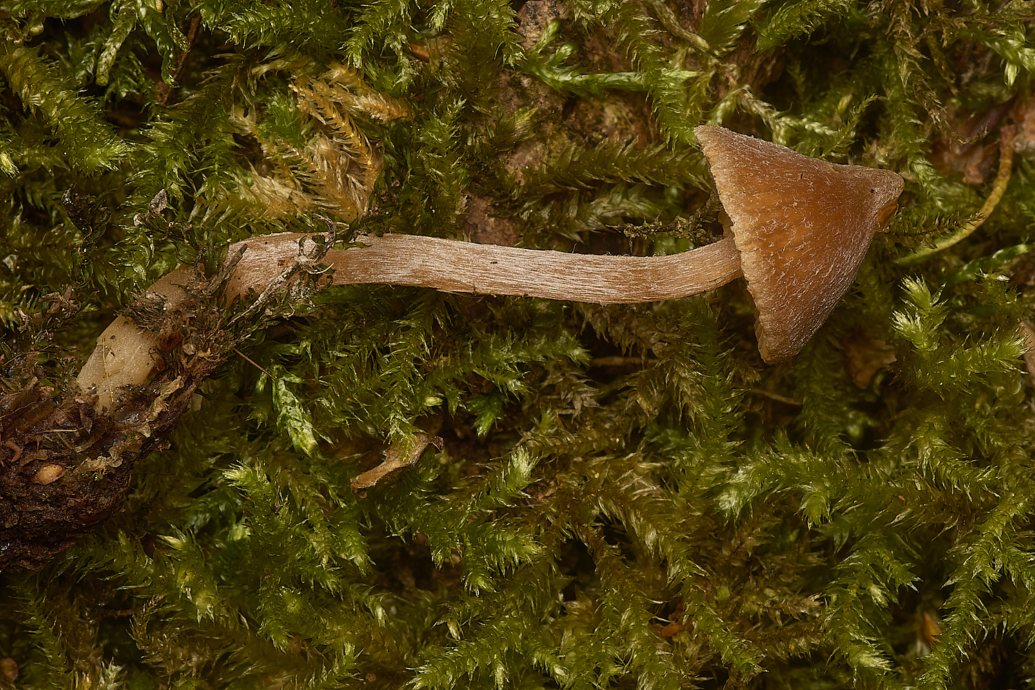
Cortinarius Sp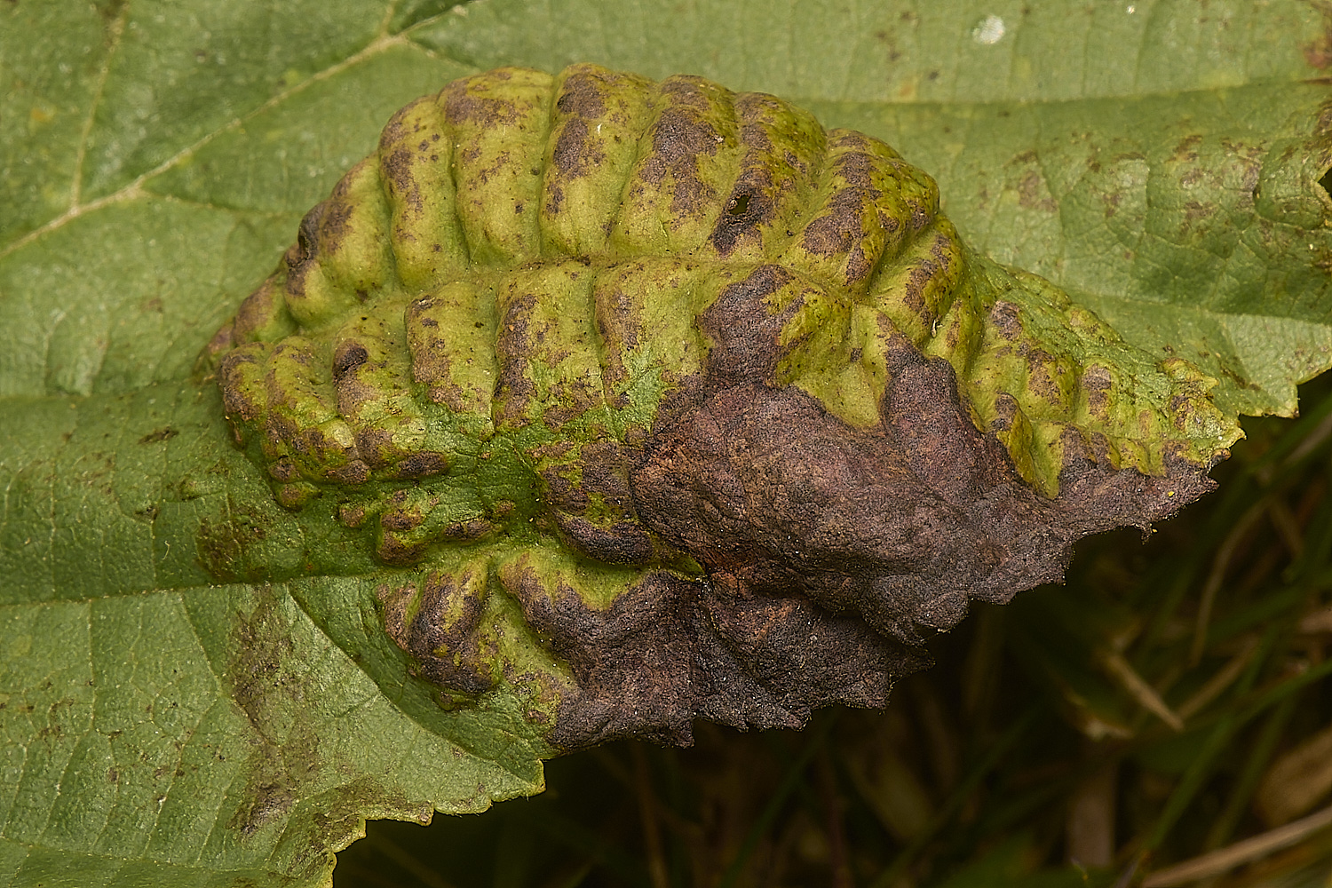
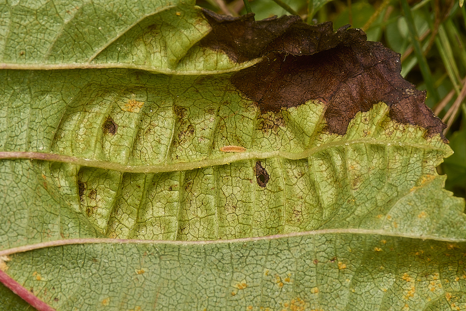
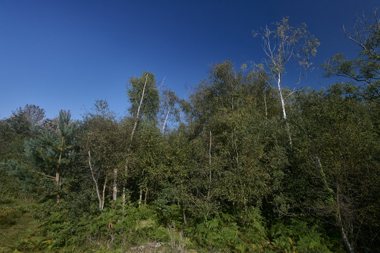
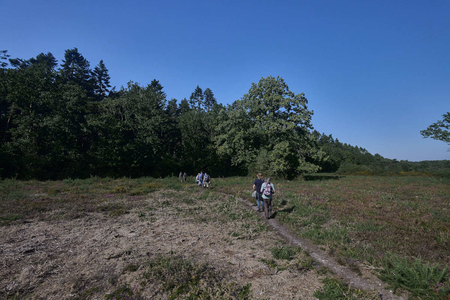
Taphrina tosquinetii causes these blisters on both side of Alder (Alnus glutinosa) leaves 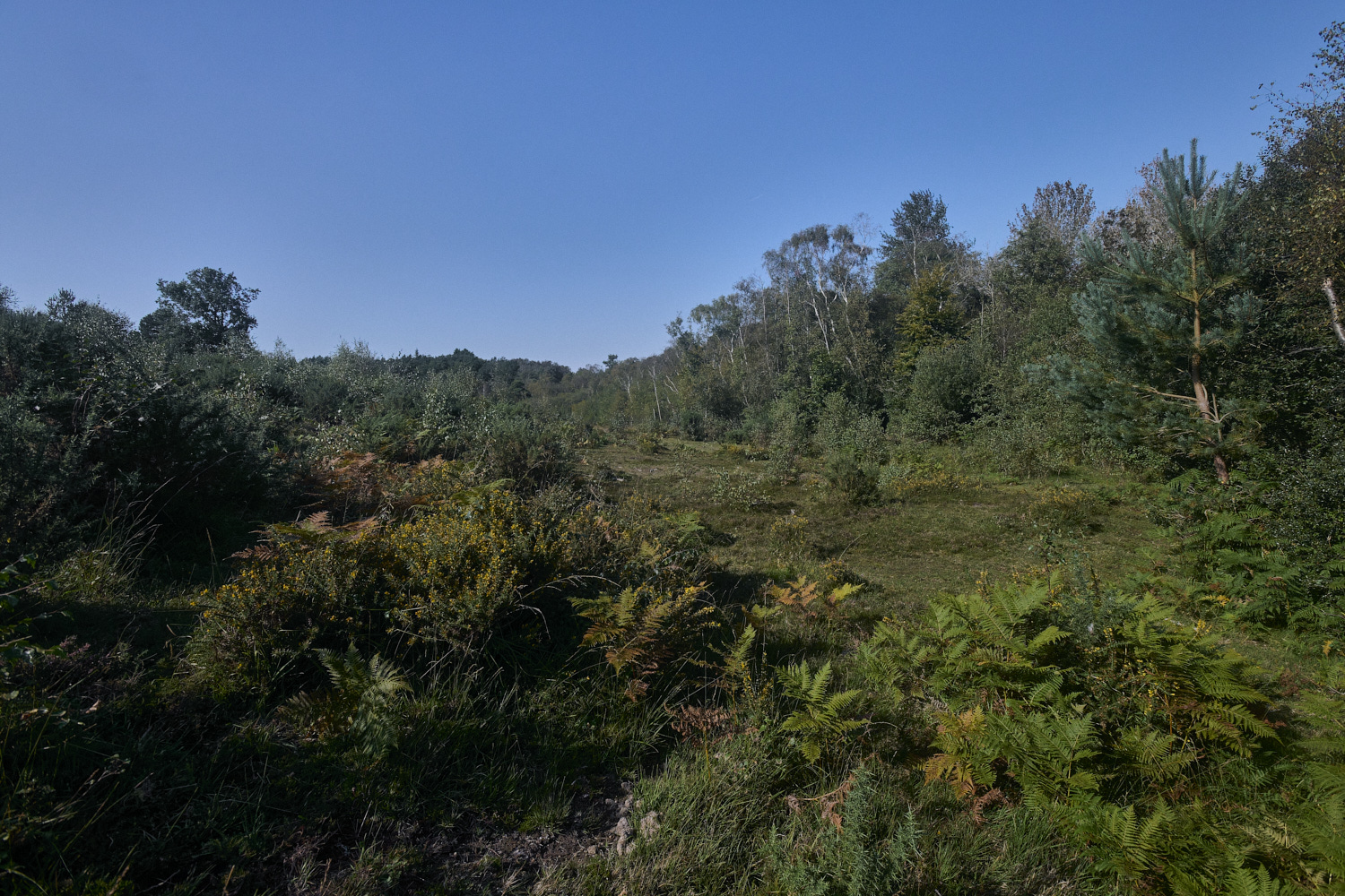
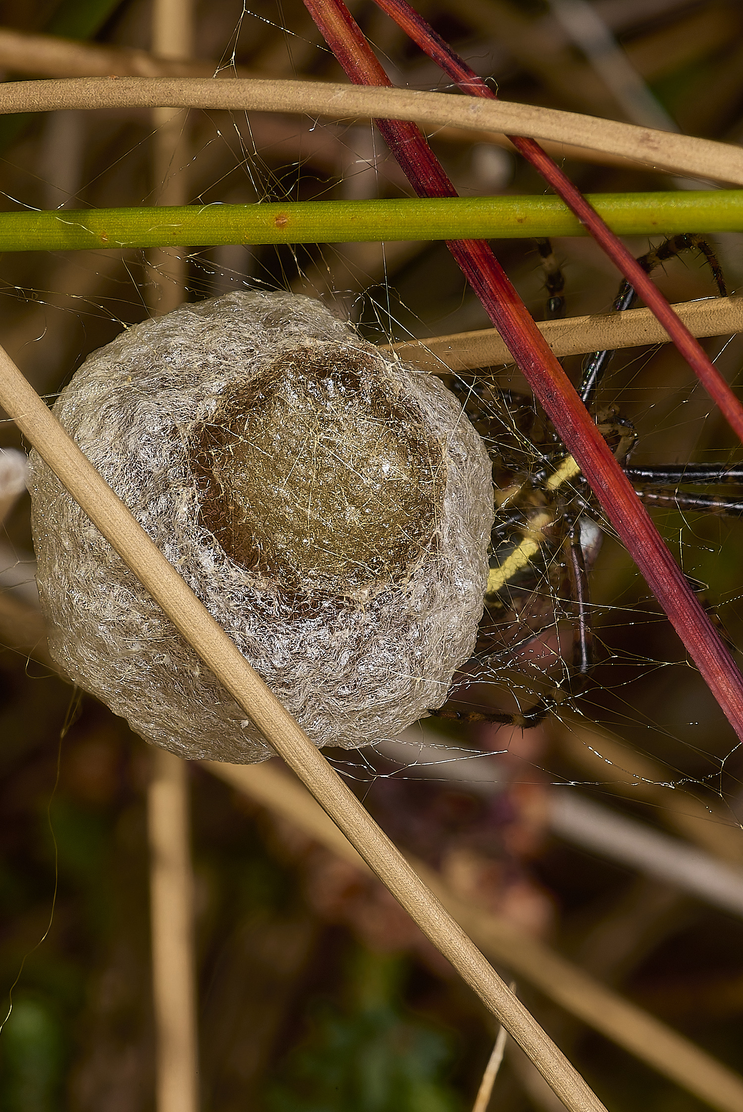
Wasp Spider egg sack (Argiope brunnechii)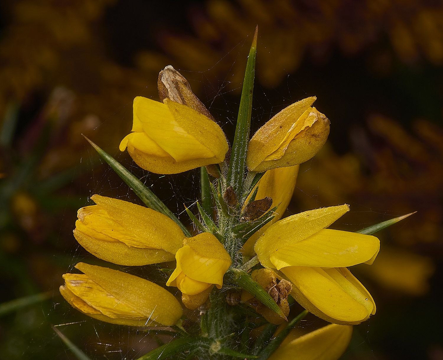
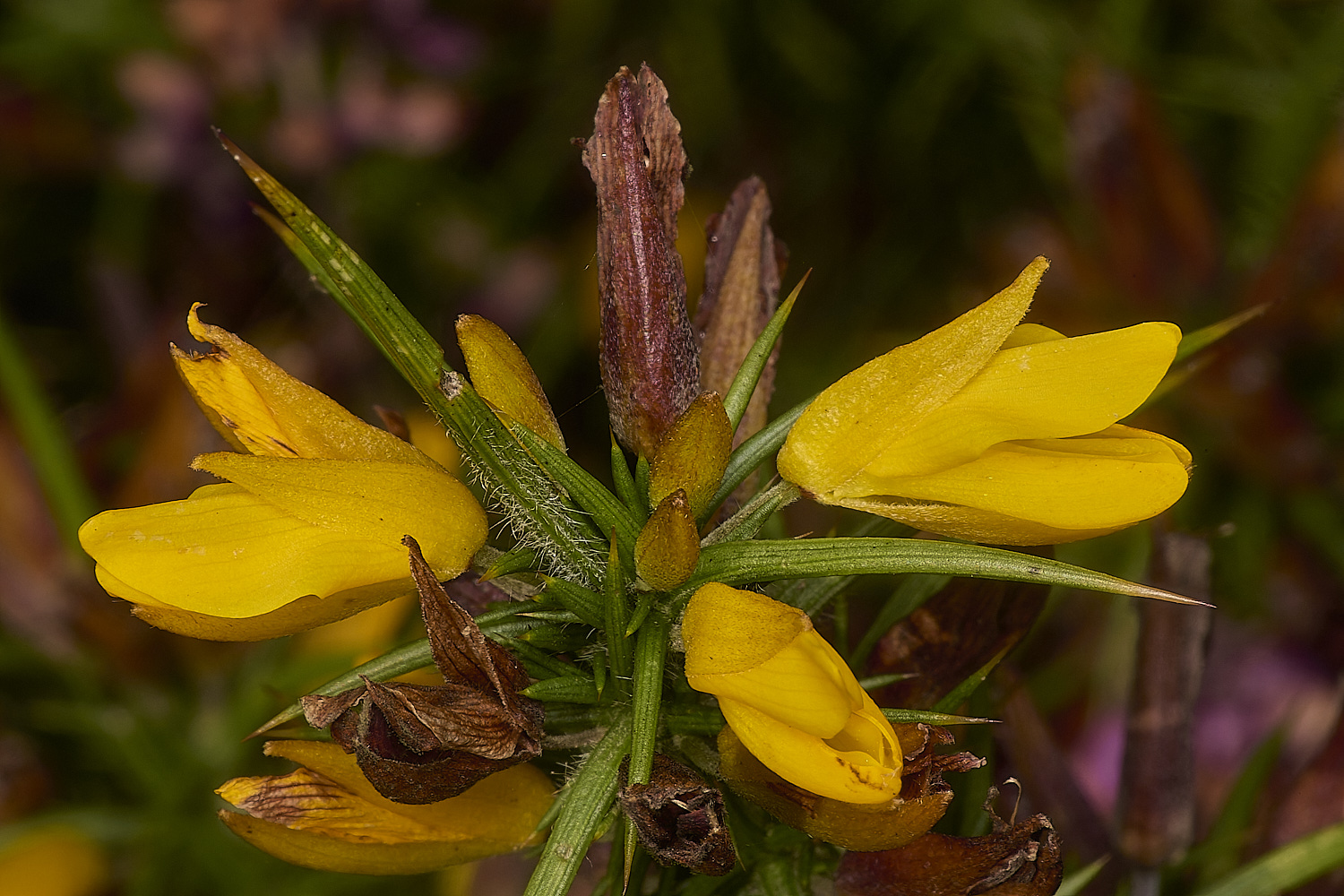
Western Gorse (Dwarf Furze) (Ulex gallii)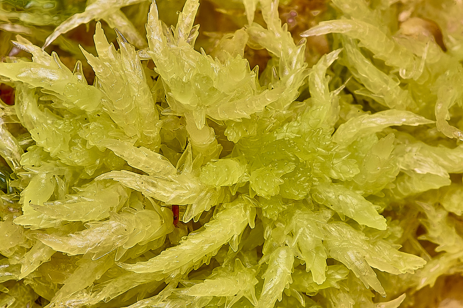
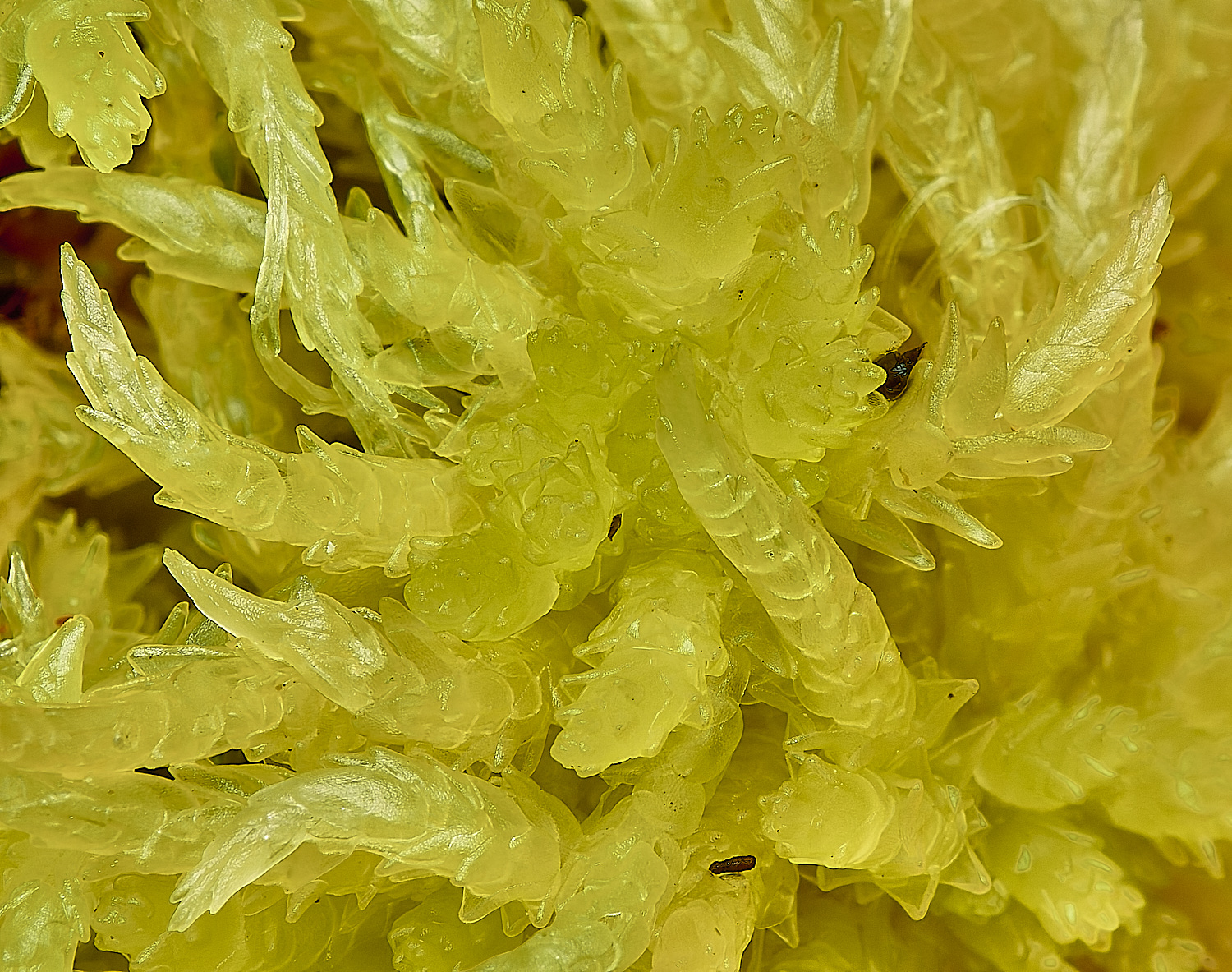
Blunt-leaved Bog-moss (Sphagnum palustre)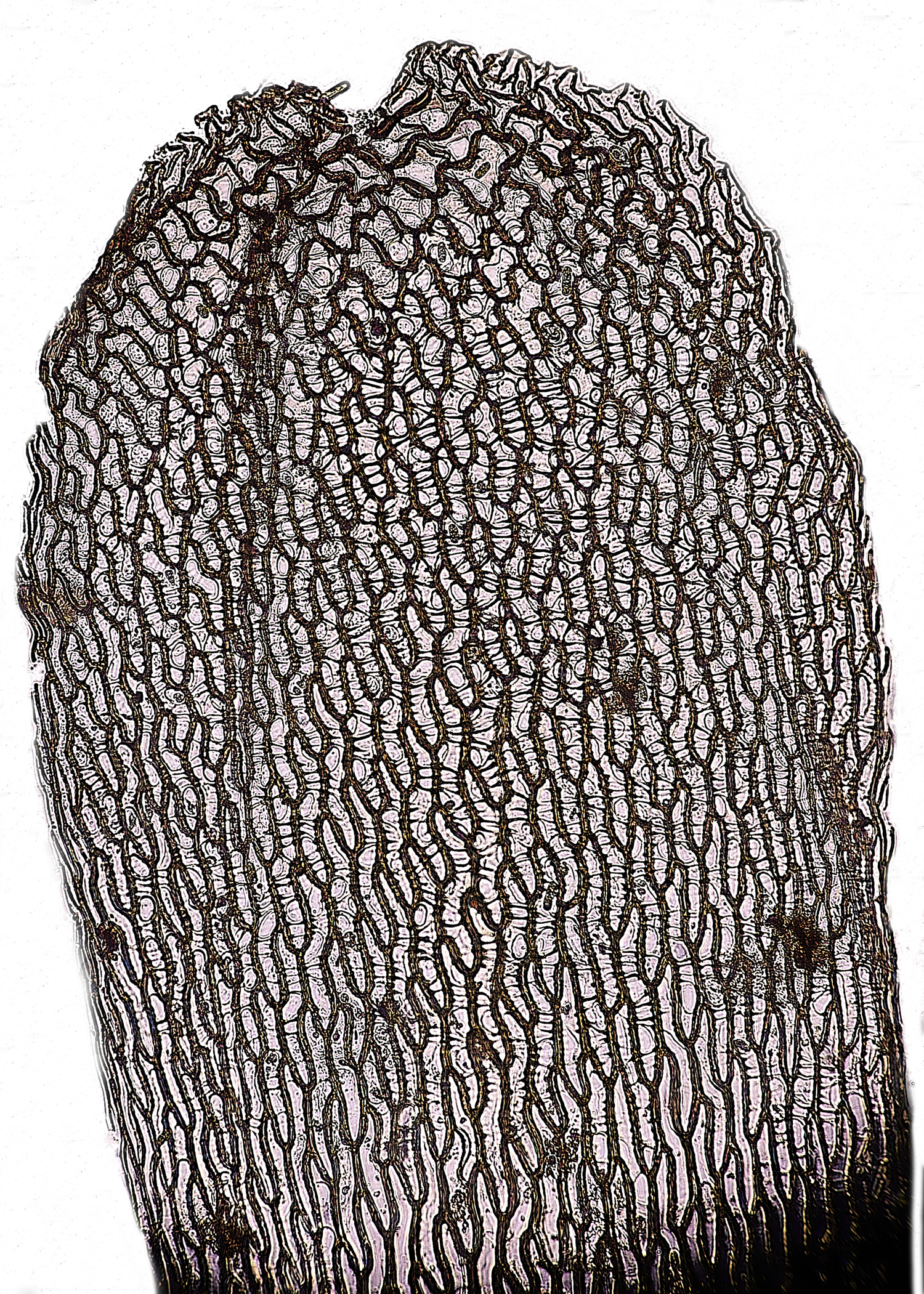
x 200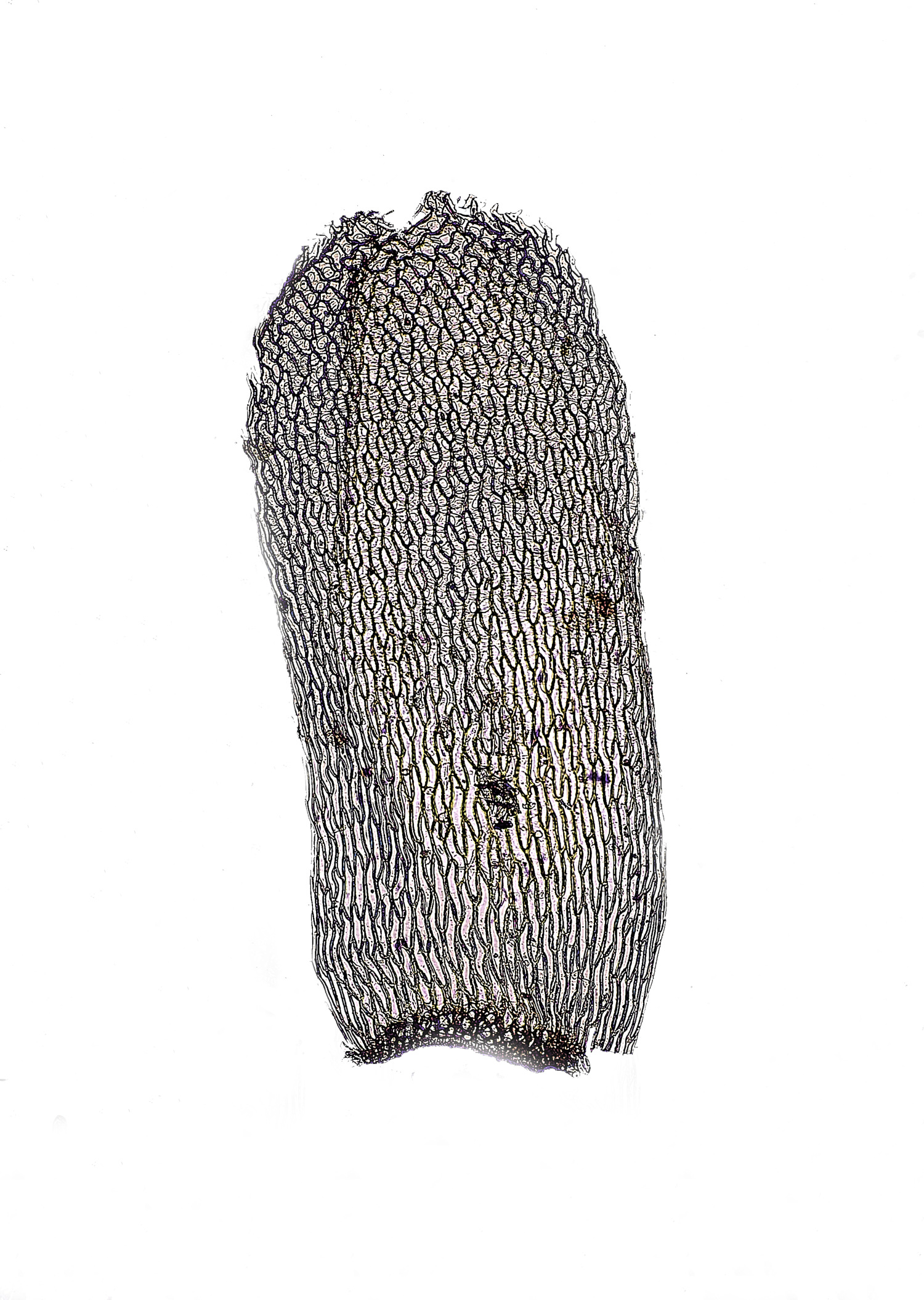
x100
Stem Leaf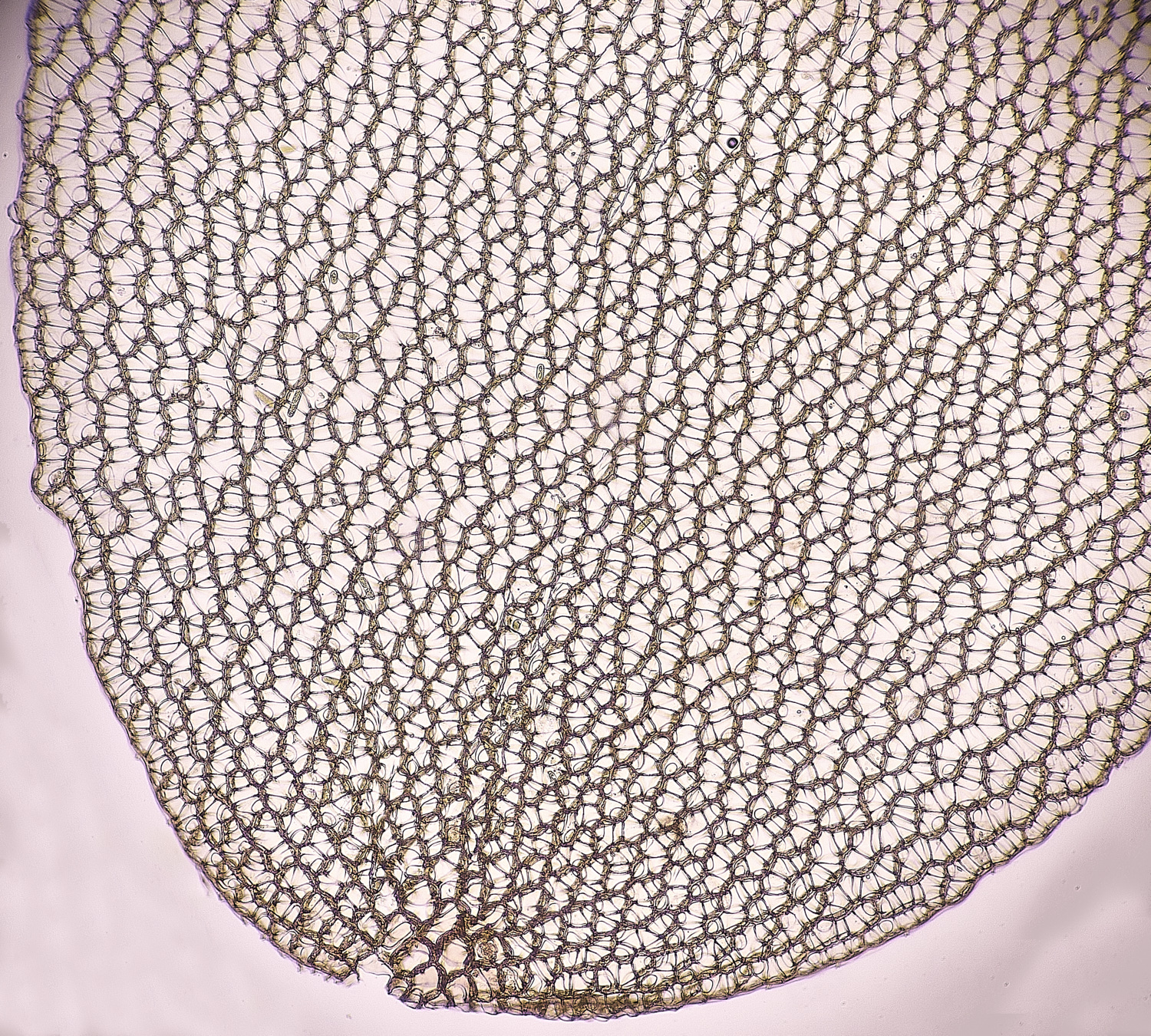
Part of branch leaf
x100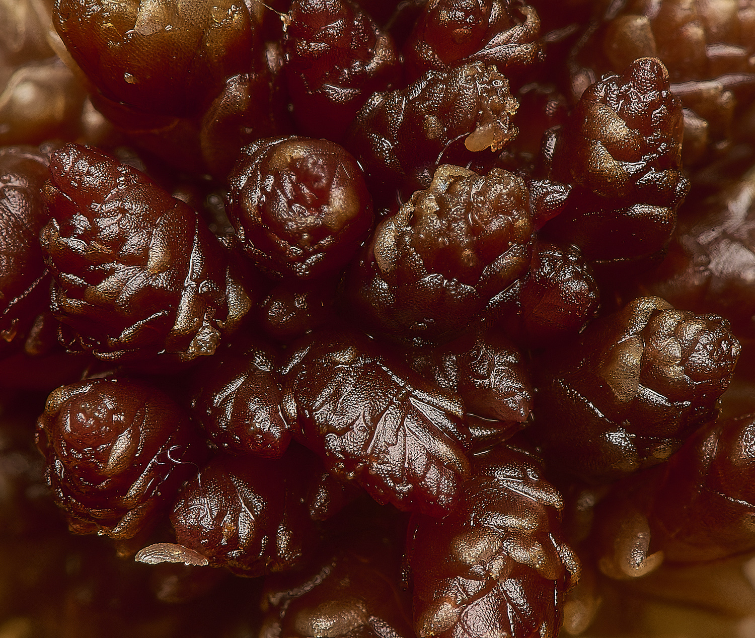
Magellanic Peatmoss (Sphagnum magellanicum agg)
It looks likely that this is Sphagnum medium but it needs to be confirmed.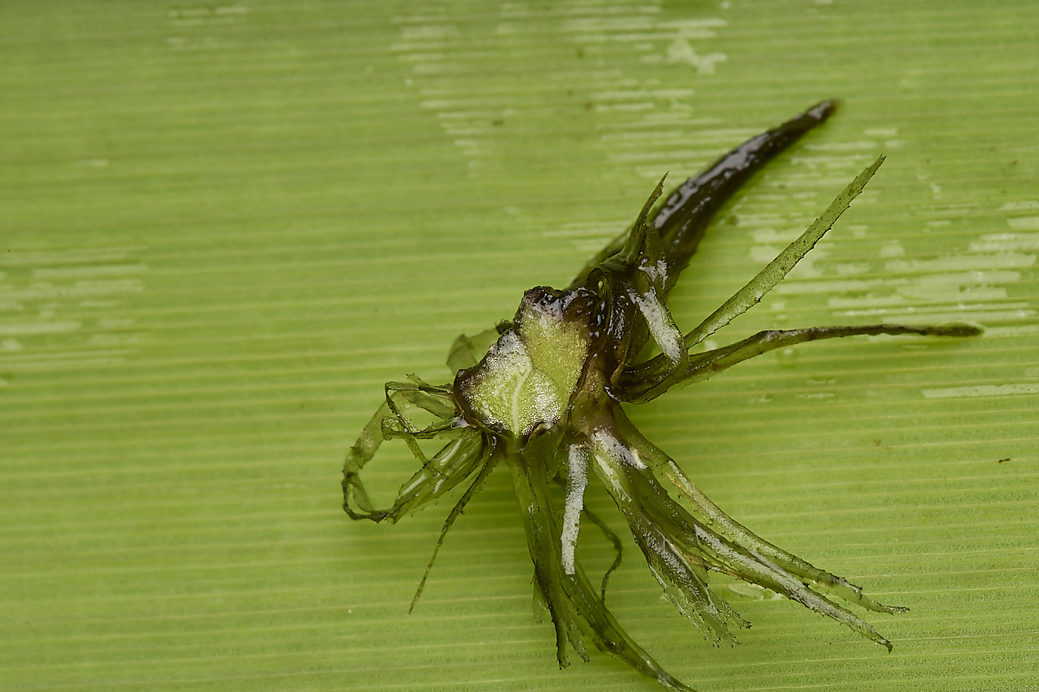
Bristle Club-rush Isolepis setacea)